 Movie
Movie Controller


+ Open data

- Basic information
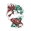
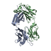
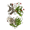
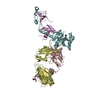
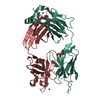
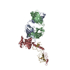
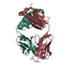
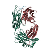
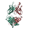
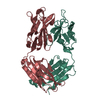
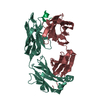
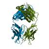
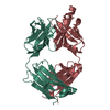
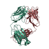
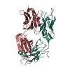
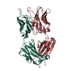
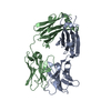
| Entry | Database: PDB / ID: 3cmo | ||||||
|---|---|---|---|---|---|---|---|
| Title | HIV neutralizing monoclonal antibody YZ18 | ||||||
 Components Components |
| ||||||
 Keywords Keywords |  IMMUNE SYSTEM / HIV-1 / monoclonal antibody IMMUNE SYSTEM / HIV-1 / monoclonal antibody | ||||||
| Function / homology |  Function and homology information Function and homology informationImmunoglobulin V-Type / Immunoglobulin V-set domain / Immunoglobulin V-set domain / Immunoglobulin/major histocompatibility complex, conserved site / Immunoglobulin subtype / Immunoglobulins and major histocompatibility complex proteins signature. / Immunoglobulin / Immunoglobulin C-Type / Immunoglobulin C1-set / Immunoglobulin C1-set domain ...Immunoglobulin V-Type / Immunoglobulin V-set domain / Immunoglobulin V-set domain / Immunoglobulin/major histocompatibility complex, conserved site / Immunoglobulin subtype / Immunoglobulins and major histocompatibility complex proteins signature. / Immunoglobulin / Immunoglobulin C-Type / Immunoglobulin C1-set / Immunoglobulin C1-set domain / Ig-like domain profile. / Immunoglobulin-like domain / Immunoglobulin-like domain superfamily / Immunoglobulins / Immunoglobulin-like fold / Immunoglobulin-like / Sandwich / Mainly BetaSimilarity search - Domain/homology | ||||||
| Biological species |  Mus musculus (house mouse) Mus musculus (house mouse) | ||||||
| Method | X-RAY DIFFRACTION / SYNCHROTRON / MOLECULAR REPLACEMENT / Resolution: 2.3 Å | ||||||
 Authors Authors | Jin, L. / Prasad, V. / Paul, S. | ||||||
 Citation Citation | Journal: TO BE PUBLISHED Title: HIV neutralizing monoclonal antibody YZ18 Authors: Jin, L. / Prasad, V. / Paul, S. | ||||||
| History |
|
- Structure visualization
| Structure viewer | Molecule: MolmilJmol/JSmol |
|---|
- Downloads & links
-Download
| PDBx/mmCIF format | 3cmo.cif.gz | 173.3 KB | Display | PDBx/mmCIF format |
|---|---|---|---|---|
| PDB format | pdb3cmo.ent.gz | 138.7 KB | Display | PDB format |
| PDBx/mmJSON format | 3cmo.json.gz | Tree view | PDBx/mmJSON format | |
| Others |  Other downloads Other downloads |
-Validation report
| Arichive directory | https://data.pdbj.org/pub/pdb/validation_reports/cm/3cmoftp://data.pdbj.org/pub/pdb/validation_reports/cm/3cmo | HTTPS FTP |
|---|
-Related structure data
| Related structure data | 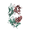 1yehS S: Starting model for refinement |
|---|---|
| Similar structure data |


-Links
 PDBj
PDBj


- Assembly
| Deposited unit | 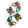
| ||||||||
|---|---|---|---|---|---|---|---|---|---|
| 1 | 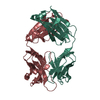
| ||||||||
| 2 | 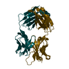
| ||||||||
| 3 |
| ||||||||
| Unit cell |
|
-Components
| #1: Antibody | Fragment antigen-binding Mass: 23264.598 Da / Num. of mol.: 2 / Source method: isolated from a natural source / Source: (natural) Mus musculus (house mouse) / Strain: hybridoma#2: Antibody | Fragment antigen-bindingMass: 23940.752 Da / Num. of mol.: 2 / Source method: isolated from a natural source / Source: (natural) Mus musculus (house mouse) / Strain: hybridoma / References: UniProt: Q6PJA7*PLUS#3: Water | ChemComp-HOH / | Water Mass: 18.015 Da / Num. of mol.: 86 / Source method: isolated from a natural source / Formula: H2O Mass: 18.015 Da / Num. of mol.: 86 / Source method: isolated from a natural source / Formula: H2O |
|---|
-Experimental details
-Experiment
| Experiment | Method: X-RAY DIFFRACTION / Number of used crystals: 1 |
|---|
- Sample preparation
| Crystal | Density Matthews: 2.36 Å3/Da / Density % sol: 47.77 % |
|---|---|
| Crystal grow | Temperature: 298 K / Method: vapor diffusion, hanging drop / pH: 5 Details: 18% PEG6000, 0.1 M sodium citrate, pH 5, VAPOR DIFFUSION, HANGING DROP, temperature 298K |
-Data collection
| Diffraction | Mean temperature: 100 K |
|---|---|
| Diffraction source | Source: SYNCHROTRON / Site: ALS  / Beamline: 4.2.2 / Wavelength: 1.24 Å / Beamline: 4.2.2 / Wavelength: 1.24 Å |
| Detector | Type: CUSTOM-MADE / Detector: CCD / Date: Dec 7, 2006 |
| Radiation | Protocol: SINGLE WAVELENGTH / Monochromatic (M) / Laue (L): M / Scattering type: x-ray |
| Radiation wavelength | Wavelength: 1.24 Å / Relative weight: 1 |
| Reflection | Resolution: 2.3→50 Å / Num. all: 39214 / Num. obs: 37445 / % possible obs: 95.5 % / Observed criterion σ(F): 0 / Observed criterion σ(I): -3 / Redundancy: 3 % / Biso Wilson estimate: 33 Å2 / Rsym value: 0.09 / Net I/σ(I): 5.9 |
| Reflection shell | Resolution: 2.3→2.38 Å / Redundancy: 2.95 % / Mean I/σ(I) obs: 1.9 / Num. unique all: 4184 / Rsym value: 0.426 / % possible all: 95 |
- Processing
| Software |
| |||||||||||||||||||||||||
|---|---|---|---|---|---|---|---|---|---|---|---|---|---|---|---|---|---|---|---|---|---|---|---|---|---|---|
| Refinement | Method to determine structure: MOLECULAR REPLACEMENT Starting model: PDB entry 1YEH Resolution: 2.3→15 Å / Isotropic thermal model: isotropic / Cross valid method: THROUGHOUT / σ(F): 0 / Stereochemistry target values: Engh & Huber
| |||||||||||||||||||||||||
| Displacement parameters | Biso mean: 39 Å2
| |||||||||||||||||||||||||
| Refinement step | Cycle: LAST / Resolution: 2.3→15 Å
| |||||||||||||||||||||||||
| Refine LS restraints |
|
