 Movie
Movie Controller


+ Open data

- Basic information
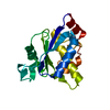
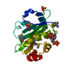
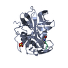
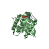
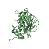
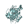
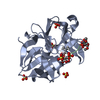
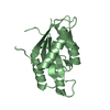
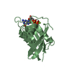
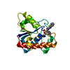
| Entry | Database: PDB / ID: 4xwx | ||||||
|---|---|---|---|---|---|---|---|
| Title | Crystal structure of the PTB domain of SHC | ||||||
 Components Components | SHC-transforming protein 1 | ||||||
 Keywords Keywords |  SIGNALING PROTEIN / phosphotyrosine binding domain / signal transduction / Structural Genomics Consortium (SGC) SIGNALING PROTEIN / phosphotyrosine binding domain / signal transduction / Structural Genomics Consortium (SGC) | ||||||
| Function / homology |  Function and homology information Function and homology informationregulation of superoxide metabolic process / positive regulation of cell proliferation in bone marrow / XBP1(S) activates chaperone genes / neurotrophin TRKA receptor binding / transmembrane receptor protein tyrosine kinase adaptor activity / Interleukin-15 signaling / Shc-EGFR complex / Interleukin-2 signaling / epidermal growth factor binding / epidermal growth factor receptor binding ...regulation of superoxide metabolic process / positive regulation of cell proliferation in bone marrow / XBP1(S) activates chaperone genes / neurotrophin TRKA receptor binding / transmembrane receptor protein tyrosine kinase adaptor activity / Interleukin-15 signaling / Shc-EGFR complex / Interleukin-2 signaling / epidermal growth factor binding / epidermal growth factor receptor binding / Signaling by ALK / Signaling by ALK fusions and activated point mutants / RET signaling / Activated NTRK3 signals through RAS / Activated NTRK2 signals through RAS / Interleukin-3, Interleukin-5 and GM-CSF signaling / SHC1 events in ERBB4 signaling / Signalling to RAS / SHC-related events triggered by IGF1R / Role of LAT2/NTAL/LAB on calcium mobilization / Signal attenuation / Interleukin receptor SHC signaling / SHC-mediated cascade:FGFR3 / MET activates RAS signaling / SHC-mediated cascade:FGFR2 / SHC-mediated cascade:FGFR4 / SHC-mediated cascade:FGFR1 / Erythropoietin activates RAS / Signaling by CSF3 (G-CSF) / Tie2 Signaling / SHC1 events in EGFR signaling / insulin-like growth factor receptor binding / phosphotyrosine residue binding / ephrin receptor binding / SHC1 events in ERBB2 signaling / Integrin signaling / negative regulation of angiogenesis / Constitutive Signaling by Overexpressed ERBB2 / FCERI mediated Ca+2 mobilization / Insulin receptor signalling cascade / insulin-like growth factor receptor signaling pathway / FCERI mediated MAPK activation / Signaling by ERBB2 TMD/JMD mutants / phospholipid binding / Constitutive Signaling by EGFRvIII / insulin receptor binding / Signaling by ERBB2 ECD mutants / epidermal growth factor receptor signaling pathway / Signaling by ERBB2 KD Mutants / cell-cell adhesion / receptor tyrosine kinase binding / cellular response to growth factor stimulus / GPER1 signaling / Signaling by CSF1 (M-CSF) in myeloid cells / Constitutive Signaling by Ligand-Responsive EGFR Cancer Variants / DAP12 signaling / insulin receptor signaling pathway / heart development / actin cytoskeleton organization / RAF/MAP kinase cascade / angiogenesis / Extra-nuclear estrogen signaling / positive regulation of MAPK cascade / positive regulation of ERK1 and ERK2 cascade / mitochondrial matrix / intracellular signal transduction / defense response to bacterium / focal adhesion / negative regulation of DNA-templated transcription / positive regulation of cell population proliferation / negative regulation of apoptotic process / positive regulation of DNA-templated transcription / plasma membrane / cytosolSimilarity search - Function | ||||||
| Biological species |  Homo sapiens (human) Homo sapiens (human) | ||||||
| Method | X-RAY DIFFRACTION / SYNCHROTRON / Resolution: 1.87 Å | ||||||
 Authors Authors | Chaikuad, A. / Tallant, C. / Krojer, T. / Dixon-Clarke, S. / von Delft, F. / Arrowsmith, C.H. / Edwards, A.M. / Bountra, C. / Knapp, S. / Structural Genomics Consortium (SGC) | ||||||
 Citation Citation | Journal: To Be Published Title: Crystal structure of the PTB domain of SHC Authors: Chaikuad, A. / Tallant, C. / Krojer, T. / Dixon-Clarke, S. / von Delft, F. / Arrowsmith, C.H. / Edwards, A.M. / Bountra, C. / Knapp, S. / Structural Genomics Consortium (SGC) | ||||||
| History |
|
- Structure visualization
| Structure viewer | Molecule: MolmilJmol/JSmol |
|---|
- Downloads & links
-Download
| PDBx/mmCIF format | 4xwx.cif.gz | 80.8 KB | Display | PDBx/mmCIF format |
|---|---|---|---|---|
| PDB format | pdb4xwx.ent.gz | 59.3 KB | Display | PDB format |
| PDBx/mmJSON format | 4xwx.json.gz | Tree view | PDBx/mmJSON format | |
| Others |  Other downloads Other downloads |
-Validation report
| Arichive directory | https://data.pdbj.org/pub/pdb/validation_reports/xw/4xwxftp://data.pdbj.org/pub/pdb/validation_reports/xw/4xwx | HTTPS FTP |
|---|
-Related structure data
| Similar structure data |
|---|
-Links
 PDBj
PDBj


- Assembly
| Deposited unit | 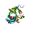
| ||||||||
|---|---|---|---|---|---|---|---|---|---|
| 1 |
| ||||||||
| Unit cell |
| ||||||||
| Components on special symmetry positions |
|
-Components
| #1: Protein | Mass: 18133.912 Da / Num. of mol.: 1 / Fragment: residues 37-201 Source method: isolated from a genetically manipulated source Details: residues 37-201 / Source: (gene. exp.) Homo sapiens (human) / Gene: SHC1, SHC, SHCA / Plasmid: pNIC28-BSA4 / Production host:  Escherichia coli BL21(DE3) (bacteria) / Variant (production host): R3 / References: UniProt: P29353 Escherichia coli BL21(DE3) (bacteria) / Variant (production host): R3 / References: UniProt: P29353 | ||||
|---|---|---|---|---|---|
| #2: Chemical | ChemComp-EDO / Ethylene glycol  Mass: 62.068 Da / Num. of mol.: 6 / Source method: obtained synthetically / Formula: C2H6O2 Mass: 62.068 Da / Num. of mol.: 6 / Source method: obtained synthetically / Formula: C2H6O2#3: Chemical | ChemComp-NA / |   Mass: 22.990 Da / Num. of mol.: 1 / Source method: obtained synthetically / Formula: Na Mass: 22.990 Da / Num. of mol.: 1 / Source method: obtained synthetically / Formula: Na#4: Water | ChemComp-HOH / | Water Mass: 18.015 Da / Num. of mol.: 101 / Source method: isolated from a natural source / Formula: H2O Mass: 18.015 Da / Num. of mol.: 101 / Source method: isolated from a natural source / Formula: H2O |
-Experimental details
-Experiment
| Experiment | Method: X-RAY DIFFRACTION |
|---|
- Sample preparation
| Crystal | Density Matthews: 2.86 Å3/Da / Density % sol: 56.98 % |
|---|---|
| Crystal grow | Temperature: 277.15 K / Method: vapor diffusion, sitting drop / pH: 7.5 Details: 0.8 M sodium/potassium tartrate, 0.1 M Hepes pH 7.5 |
-Data collection
| Diffraction | Mean temperature: 100 K |
|---|---|
| Diffraction source | Source: SYNCHROTRON / Site: Diamond  / Beamline: I04 / Wavelength: 0.97949 Å / Beamline: I04 / Wavelength: 0.97949 Å |
| Detector | Type: PSI PILATUS 6M / Detector: PIXEL / Date: Apr 14, 2014 |
| Radiation | Monochromator: Si (111) double crystal monochromator / Protocol: SINGLE WAVELENGTH / Monochromatic (M) / Laue (L): M / Scattering type: x-ray |
| Radiation wavelength | Wavelength: 0.97949 Å / Relative weight: 1 |
| Reflection | Resolution: 1.87→28.98 Å / Num. all: 17585 / Num. obs: 17581 / % possible obs: 99.9 % / Redundancy: 5.3 % / Biso Wilson estimate: 35.1 Å2 / Rmerge(I) obs: 0.06 / Net I/σ(I): 13.4 |
| Reflection shell | Resolution: 1.87→1.97 Å / Redundancy: 5.3 % / Rmerge(I) obs: 0.823 / Mean I/σ(I) obs: 2.1 / % possible all: 100 |
- Processing
| Software |
| ||||||||||||||||||||||||||||||||||||||||||||||||||||||||||||||||||||||||||||||||||||||||||||||||||||||||||||||||||||||||||||||||||||||||||||||||||||||||||||||||||||||||||||||||||||||
|---|---|---|---|---|---|---|---|---|---|---|---|---|---|---|---|---|---|---|---|---|---|---|---|---|---|---|---|---|---|---|---|---|---|---|---|---|---|---|---|---|---|---|---|---|---|---|---|---|---|---|---|---|---|---|---|---|---|---|---|---|---|---|---|---|---|---|---|---|---|---|---|---|---|---|---|---|---|---|---|---|---|---|---|---|---|---|---|---|---|---|---|---|---|---|---|---|---|---|---|---|---|---|---|---|---|---|---|---|---|---|---|---|---|---|---|---|---|---|---|---|---|---|---|---|---|---|---|---|---|---|---|---|---|---|---|---|---|---|---|---|---|---|---|---|---|---|---|---|---|---|---|---|---|---|---|---|---|---|---|---|---|---|---|---|---|---|---|---|---|---|---|---|---|---|---|---|---|---|---|---|---|---|---|
| Refinement | Resolution: 1.87→28.98 Å / Cor.coef. Fo:Fc: 0.975 / Cor.coef. Fo:Fc free: 0.968 / SU B: 7.053 / SU ML: 0.098 / Cross valid method: THROUGHOUT / ESU R: 0.11 / ESU R Free: 0.103 / Stereochemistry target values: MAXIMUM LIKELIHOOD / Details: HYDROGENS HAVE BEEN ADDED IN THE RIDING POSITIONS
| ||||||||||||||||||||||||||||||||||||||||||||||||||||||||||||||||||||||||||||||||||||||||||||||||||||||||||||||||||||||||||||||||||||||||||||||||||||||||||||||||||||||||||||||||||||||
| Solvent computation | Ion probe radii: 0.8 Å / Shrinkage radii: 0.8 Å / VDW probe radii: 1.2 Å / Solvent model: MASK | ||||||||||||||||||||||||||||||||||||||||||||||||||||||||||||||||||||||||||||||||||||||||||||||||||||||||||||||||||||||||||||||||||||||||||||||||||||||||||||||||||||||||||||||||||||||
| Displacement parameters | Biso mean: 47.944 Å2
| ||||||||||||||||||||||||||||||||||||||||||||||||||||||||||||||||||||||||||||||||||||||||||||||||||||||||||||||||||||||||||||||||||||||||||||||||||||||||||||||||||||||||||||||||||||||
| Refine analyze | Luzzati coordinate error obs: 0.253 Å | ||||||||||||||||||||||||||||||||||||||||||||||||||||||||||||||||||||||||||||||||||||||||||||||||||||||||||||||||||||||||||||||||||||||||||||||||||||||||||||||||||||||||||||||||||||||
| Refinement step | Cycle: 1 / Resolution: 1.87→28.98 Å
| ||||||||||||||||||||||||||||||||||||||||||||||||||||||||||||||||||||||||||||||||||||||||||||||||||||||||||||||||||||||||||||||||||||||||||||||||||||||||||||||||||||||||||||||||||||||
| Refine LS restraints |
|