 Movie
Movie Controller

[English] 日本語
 Yorodumi
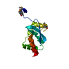
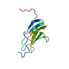
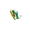
- PDB-4xtb: Crystal structure of the N-terminal domain of the human mitochond... -
+ Open data

- Basic information
| Entry | Database: PDB / ID: 4xtb | ||||||
|---|---|---|---|---|---|---|---|
| Title | Crystal structure of the N-terminal domain of the human mitochondrial calcium uniporter | ||||||
 Components Components | Calcium uniporter protein, mitochondrial | ||||||
 Keywords Keywords |  TRANSPORT PROTEIN / Calcium channel / Membrane protein / Mitochondria TRANSPORT PROTEIN / Calcium channel / Membrane protein / Mitochondria | ||||||
| Function / homology |  Function and homology informationuniporter activity / Processing of SMDT1 / mitochondrial calcium ion transmembrane transport / uniplex complex / Mitochondrial calcium ion transport / calcium import into the mitochondrion / positive regulation of mitochondrial calcium ion concentration / mitochondrial calcium ion homeostasis / positive regulation of neutrophil chemotaxis / positive regulation of mitochondrial fission ...uniporter activity / Processing of SMDT1 / mitochondrial calcium ion transmembrane transport / uniplex complex / Mitochondrial calcium ion transport / calcium import into the mitochondrion / positive regulation of mitochondrial calcium ion concentration / mitochondrial calcium ion homeostasis / positive regulation of neutrophil chemotaxis / positive regulation of mitochondrial fission / calcium channel complex / calcium-mediated signaling / calcium channel activity / positive regulation of insulin secretion / protein complex oligomerization / glucose homeostasis / mitochondrial inner membrane / mitochondrion / identical protein binding Function and homology informationuniporter activity / Processing of SMDT1 / mitochondrial calcium ion transmembrane transport / uniplex complex / Mitochondrial calcium ion transport / calcium import into the mitochondrion / positive regulation of mitochondrial calcium ion concentration / mitochondrial calcium ion homeostasis / positive regulation of neutrophil chemotaxis / positive regulation of mitochondrial fission ...uniporter activity / Processing of SMDT1 / mitochondrial calcium ion transmembrane transport / uniplex complex / Mitochondrial calcium ion transport / calcium import into the mitochondrion / positive regulation of mitochondrial calcium ion concentration / mitochondrial calcium ion homeostasis / positive regulation of neutrophil chemotaxis / positive regulation of mitochondrial fission / calcium channel complex / calcium-mediated signaling / calcium channel activity / positive regulation of insulin secretion / protein complex oligomerization / glucose homeostasis / mitochondrial inner membrane / mitochondrion / identical protein bindingSimilarity search - Function | ||||||
| Biological species |  Homo sapiens (human) Homo sapiens (human) | ||||||
| Method | X-RAY DIFFRACTION / SYNCHROTRON / MOLECULAR REPLACEMENT / Resolution: 1.5 Å | ||||||
 Authors Authors | Lee, Y. / Min, C.K. / Kim, T.G. / Song, H.K. / Lim, Y. / Kim, D. / Shin, K. / Kang, M. / Kang, J.Y. / Youn, H.-S. ...Lee, Y. / Min, C.K. / Kim, T.G. / Song, H.K. / Lim, Y. / Kim, D. / Shin, K. / Kang, M. / Kang, J.Y. / Youn, H.-S. / Lee, J.-G. / An, J.Y. / Park, K.R. / Lim, J.J. / Kim, J.H. / Kim, J.H. / Park, Z.Y. / Kim, Y.-S. / Wang, J. / Kim, D.H. / Eom, S.H. | ||||||
 Citation Citation | Journal: Embo Rep. / Year: 2015 Title: Structure and function of the N-terminal domain of the human mitochondrial calcium uniporter. Authors: Lee, Y. / Min, C.K. / Kim, T.G. / Song, H.K. / Lim, Y. / Kim, D. / Shin, K. / Kang, M. / Kang, J.Y. / Youn, H.S. / Lee, J.G. / An, J.Y. / Park, K.R. / Lim, J.J. / Kim, J.H. / Kim, J.H. / ...Authors: Lee, Y. / Min, C.K. / Kim, T.G. / Song, H.K. / Lim, Y. / Kim, D. / Shin, K. / Kang, M. / Kang, J.Y. / Youn, H.S. / Lee, J.G. / An, J.Y. / Park, K.R. / Lim, J.J. / Kim, J.H. / Kim, J.H. / Park, Z.Y. / Kim, Y.S. / Wang, J. / Kim, D.H. / Eom, S.H. | ||||||
| History |
|
- Structure visualization
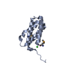
| Structure viewer | Molecule: MolmilJmol/JSmol |
|---|
- Downloads & links
-Download
| PDBx/mmCIF format | 4xtb.cif.gz | 107.3 KB | Display | PDBx/mmCIF format |
|---|---|---|---|---|
| PDB format | pdb4xtb.ent.gz | 82.4 KB | Display | PDB format |
| PDBx/mmJSON format | 4xtb.json.gz | Tree view | PDBx/mmJSON format | |
| Others |  Other downloads Other downloads |
-Validation report
| Arichive directory | https://data.pdbj.org/pub/pdb/validation_reports/xt/4xtbftp://data.pdbj.org/pub/pdb/validation_reports/xt/4xtb | HTTPS FTP |
|---|
-Related structure data
| Related structure data | 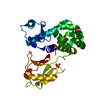 4xsjSC 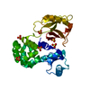 5bz6C S: Starting model for refinement C: citing same article ( |
|---|---|
| Similar structure data |


-Links
 PDBj
PDBj- Assembly
| Deposited unit | 
| ||||||||
|---|---|---|---|---|---|---|---|---|---|
| 1 |
| ||||||||
| Unit cell |
|
-Components
| #1: Protein | Mass: 14085.148 Da / Num. of mol.: 1 / Fragment: N-terminal domain (UNP RESIDUES 75-185) Source method: isolated from a genetically manipulated source Source: (gene. exp.) Homo sapiens (human) / Gene: MCU, C10orf42, CCDC109A / Production host:  Escherichia coli (E. coli) / References: UniProt: Q8NE86 Escherichia coli (E. coli) / References: UniProt: Q8NE86 |
|---|---|
| #2: Chemical | ChemComp-PG4 / Polyethylene glycol  Mass: 194.226 Da / Num. of mol.: 1 / Source method: obtained synthetically / Formula: C8H18O5 / Comment: precipitant*YM Mass: 194.226 Da / Num. of mol.: 1 / Source method: obtained synthetically / Formula: C8H18O5 / Comment: precipitant*YM |
| #3: Water | ChemComp-HOH / Water Mass: 18.015 Da / Num. of mol.: 143 / Source method: isolated from a natural source / Formula: H2O Mass: 18.015 Da / Num. of mol.: 143 / Source method: isolated from a natural source / Formula: H2O |
-Experimental details
-Experiment
| Experiment | Method: X-RAY DIFFRACTION |
|---|
- Sample preparation
| Crystal | Density Matthews: 2.17 Å3/Da / Density % sol: 43.37 % |
|---|---|
| Crystal grow | Temperature: 293 K / Method: vapor diffusion, hanging drop / pH: 8 Details: 1.55 M lithium sulphate 0.1 M Bis-Tris propane (pH 8.0) PH range: 7.0-8.0 |
-Data collection
| Diffraction | Mean temperature: 100 K |
|---|---|
| Diffraction source | Source: SYNCHROTRON / Site: PAL/PLS  / Beamline: 7A (6B, 6C1) / Wavelength: 0.9793 Å / Beamline: 7A (6B, 6C1) / Wavelength: 0.9793 Å |
| Detector | Type: ADSC QUANTUM 270 / Detector: CCD / Date: Apr 12, 2014 |
| Radiation | Protocol: SINGLE WAVELENGTH / Monochromatic (M) / Laue (L): M / Scattering type: x-ray |
| Radiation wavelength | Wavelength: 0.9793 Å / Relative weight: 1 |
| Reflection | Resolution: 1.37→100 Å / Num. obs: 23888 / % possible obs: 99.4 % / Redundancy: 5 % / Net I/σ(I): 14.3 |
| Reflection shell | Resolution: 1.37→1.39 Å / Mean I/σ(I) obs: 1.3 / % possible all: 99.6 |
- Processing
| Software |
| ||||||||||||||||||||||||||||||||||||||||||||||||||||||||||||||||||||||||||||||||||||||||||||||||||||||||||||||||||||||||||||||||||||||||||||||||||||||||||||||||||||||||||||||||||||||
|---|---|---|---|---|---|---|---|---|---|---|---|---|---|---|---|---|---|---|---|---|---|---|---|---|---|---|---|---|---|---|---|---|---|---|---|---|---|---|---|---|---|---|---|---|---|---|---|---|---|---|---|---|---|---|---|---|---|---|---|---|---|---|---|---|---|---|---|---|---|---|---|---|---|---|---|---|---|---|---|---|---|---|---|---|---|---|---|---|---|---|---|---|---|---|---|---|---|---|---|---|---|---|---|---|---|---|---|---|---|---|---|---|---|---|---|---|---|---|---|---|---|---|---|---|---|---|---|---|---|---|---|---|---|---|---|---|---|---|---|---|---|---|---|---|---|---|---|---|---|---|---|---|---|---|---|---|---|---|---|---|---|---|---|---|---|---|---|---|---|---|---|---|---|---|---|---|---|---|---|---|---|---|---|
| Refinement | Method to determine structure: MOLECULAR REPLACEMENT Starting model: 4XSJ Resolution: 1.5→48.03 Å / Cor.coef. Fo:Fc: 0.968 / Cor.coef. Fo:Fc free: 0.953 / SU B: 2.308 / SU ML: 0.039 / Cross valid method: THROUGHOUT / ESU R: 0.076 / ESU R Free: 0.066 / Stereochemistry target values: MAXIMUM LIKELIHOOD / Details: HYDROGENS HAVE BEEN ADDED IN THE RIDING POSITIONS
| ||||||||||||||||||||||||||||||||||||||||||||||||||||||||||||||||||||||||||||||||||||||||||||||||||||||||||||||||||||||||||||||||||||||||||||||||||||||||||||||||||||||||||||||||||||||
| Solvent computation | Ion probe radii: 0.8 Å / Shrinkage radii: 0.8 Å / VDW probe radii: 1.2 Å / Solvent model: MASK | ||||||||||||||||||||||||||||||||||||||||||||||||||||||||||||||||||||||||||||||||||||||||||||||||||||||||||||||||||||||||||||||||||||||||||||||||||||||||||||||||||||||||||||||||||||||
| Displacement parameters | Biso mean: 20.218 Å2
| ||||||||||||||||||||||||||||||||||||||||||||||||||||||||||||||||||||||||||||||||||||||||||||||||||||||||||||||||||||||||||||||||||||||||||||||||||||||||||||||||||||||||||||||||||||||
| Refinement step | Cycle: LAST / Resolution: 1.5→48.03 Å
| ||||||||||||||||||||||||||||||||||||||||||||||||||||||||||||||||||||||||||||||||||||||||||||||||||||||||||||||||||||||||||||||||||||||||||||||||||||||||||||||||||||||||||||||||||||||
| Refine LS restraints |
|