 Movie
Movie Controller


+ Open data

- Basic information
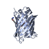
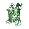
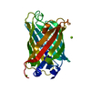
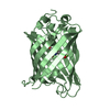
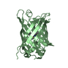
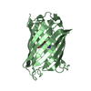
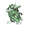
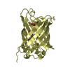
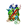
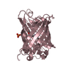
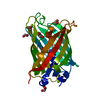
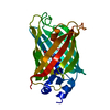
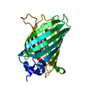
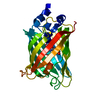
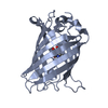
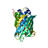
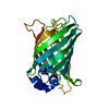
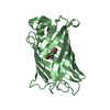
| Entry | Database: PDB / ID: 4xov | |||||||||
|---|---|---|---|---|---|---|---|---|---|---|
| Title | Structure of rsGreen0.7 in the green-off-state | |||||||||
 Components Components | rsGreen0.7 | |||||||||
 Keywords Keywords |  FLUORESCENT PROTEIN / Green Fluorescent Proteins / Reversible photoswitchablility FLUORESCENT PROTEIN / Green Fluorescent Proteins / Reversible photoswitchablility | |||||||||
| Function / homology |  Function and homology information Function and homology information | |||||||||
| Biological species |   Aequorea victoria (jellyfish) Aequorea victoria (jellyfish) | |||||||||
| Method | X-RAY DIFFRACTION / SYNCHROTRON / MOLECULAR REPLACEMENT / Resolution: 1.2 Å | |||||||||
 Authors Authors | De Zitter, E. / Van Meervelt, L. | |||||||||
| Funding support |  Belgium, 1items Belgium, 1items
| |||||||||
 Citation Citation | Journal: Acs Nano / Year: 2015 Title: Expression-Enhanced Fluorescent Proteins Based on Enhanced Green Fluorescent Protein for Super-resolution Microscopy. Authors: Duwe, S. / De Zitter, E. / Gielen, V. / Moeyaert, B. / Vandenberg, W. / Grotjohann, T. / Clays, K. / Jakobs, S. / Van Meervelt, L. / Dedecker, P. | |||||||||
| History |
|
- Structure visualization
| Structure viewer | Molecule: MolmilJmol/JSmol |
|---|
- Downloads & links
-Download
| PDBx/mmCIF format | 4xov.cif.gz | 156.4 KB | Display | PDBx/mmCIF format |
|---|---|---|---|---|
| PDB format | pdb4xov.ent.gz | 123.6 KB | Display | PDB format |
| PDBx/mmJSON format | 4xov.json.gz | Tree view | PDBx/mmJSON format | |
| Others |  Other downloads Other downloads |
-Validation report
| Arichive directory | https://data.pdbj.org/pub/pdb/validation_reports/xo/4xovftp://data.pdbj.org/pub/pdb/validation_reports/xo/4xov | HTTPS FTP |
|---|
-Related structure data


-Links
 PDBj
PDBj

- Assembly
| Deposited unit | 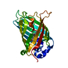
| ||||||||
|---|---|---|---|---|---|---|---|---|---|
| 1 |
| ||||||||
| Unit cell |
|
-Components
| #1: Protein | Mass: 30669.439 Da / Num. of mol.: 1 Source method: isolated from a genetically manipulated source Source: (gene. exp.) Aequorea victoria (jellyfish) / Gene: GFP / Production host:  Escherichia coli (E. coli) / Strain (production host): JM109 / References: UniProt: P42212 Escherichia coli (E. coli) / Strain (production host): JM109 / References: UniProt: P42212 |
|---|---|
| #2: Water | ChemComp-HOH / Water Mass: 18.015 Da / Num. of mol.: 291 / Source method: isolated from a natural source / Formula: H2O Mass: 18.015 Da / Num. of mol.: 291 / Source method: isolated from a natural source / Formula: H2O |
-Experimental details
-Experiment
| Experiment | Method: X-RAY DIFFRACTION / Number of used crystals: 1 |
|---|
- Sample preparation
| Crystal | Density % sol: 27.24 % |
|---|---|
| Crystal grow | Temperature: 289 K / Method: vapor diffusion, sitting drop / pH: 7.4 / Details: 0.15 M NaBr, 30% PEG 2000 MME |
-Data collection
| Diffraction | Mean temperature: 100 K |
|---|---|
| Diffraction source | Source: SYNCHROTRON / Site: SOLEIL  / Beamline: PROXIMA 1 / Wavelength: 0.8856 Å / Beamline: PROXIMA 1 / Wavelength: 0.8856 Å |
| Detector | Type: DECTRIS PILATUS 6M / Detector: PIXEL / Date: Sep 11, 2014 |
| Radiation | Protocol: SINGLE WAVELENGTH / Monochromatic (M) / Laue (L): M / Scattering type: x-ray |
| Radiation wavelength | Wavelength: 0.8856 Å / Relative weight: 1 |
| Reflection | Resolution: 1.2→40.04 Å / Num. obs: 125686 / % possible obs: 100 % / Redundancy: 3.8 % / Biso Wilson estimate: 13.81 Å2 / Rmerge(I) obs: 0.157 / Net I/σ(I): 4.9 |
| Reflection shell | Resolution: 1.2→1.24 Å / Redundancy: 3.7 % / Rmerge(I) obs: 0.613 / Mean I/σ(I) obs: 1.4 / Num. unique all: 12598 / % possible all: 100 |
- Processing
| Software |
| ||||||||||||||||||||||||||||||||||||||||||||||||||||||||||||||||||||||||||||||||||||||||||||||||||||||||||||||||||||||||||||||||||||||||||||||||||||||||||||||||||||||||
|---|---|---|---|---|---|---|---|---|---|---|---|---|---|---|---|---|---|---|---|---|---|---|---|---|---|---|---|---|---|---|---|---|---|---|---|---|---|---|---|---|---|---|---|---|---|---|---|---|---|---|---|---|---|---|---|---|---|---|---|---|---|---|---|---|---|---|---|---|---|---|---|---|---|---|---|---|---|---|---|---|---|---|---|---|---|---|---|---|---|---|---|---|---|---|---|---|---|---|---|---|---|---|---|---|---|---|---|---|---|---|---|---|---|---|---|---|---|---|---|---|---|---|---|---|---|---|---|---|---|---|---|---|---|---|---|---|---|---|---|---|---|---|---|---|---|---|---|---|---|---|---|---|---|---|---|---|---|---|---|---|---|---|---|---|---|---|---|---|---|
| Refinement | Method to determine structure: MOLECULAR REPLACEMENT / Resolution: 1.2→40.038 Å / SU ML: 0.11 / Cross valid method: FREE R-VALUE / σ(F): 1.38 / Phase error: 15.37 / Stereochemistry target values: ML
| ||||||||||||||||||||||||||||||||||||||||||||||||||||||||||||||||||||||||||||||||||||||||||||||||||||||||||||||||||||||||||||||||||||||||||||||||||||||||||||||||||||||||
| Solvent computation | Shrinkage radii: 0.9 Å / VDW probe radii: 1.11 Å / Solvent model: FLAT BULK SOLVENT MODEL | ||||||||||||||||||||||||||||||||||||||||||||||||||||||||||||||||||||||||||||||||||||||||||||||||||||||||||||||||||||||||||||||||||||||||||||||||||||||||||||||||||||||||
| Displacement parameters | Biso max: 141.56 Å2 / Biso mean: 21.8065 Å2 / Biso min: 4.36 Å2 | ||||||||||||||||||||||||||||||||||||||||||||||||||||||||||||||||||||||||||||||||||||||||||||||||||||||||||||||||||||||||||||||||||||||||||||||||||||||||||||||||||||||||
| Refinement step | Cycle: final / Resolution: 1.2→40.038 Å
| ||||||||||||||||||||||||||||||||||||||||||||||||||||||||||||||||||||||||||||||||||||||||||||||||||||||||||||||||||||||||||||||||||||||||||||||||||||||||||||||||||||||||
| Refine LS restraints |
| ||||||||||||||||||||||||||||||||||||||||||||||||||||||||||||||||||||||||||||||||||||||||||||||||||||||||||||||||||||||||||||||||||||||||||||||||||||||||||||||||||||||||
| LS refinement shell | Refine-ID: X-RAY DIFFRACTION / Total num. of bins used: 27 / % reflection obs: 100 %
|
