[English] 日本語
Yorodumi
- PDB-5hzo: GFP mutant S205G -

+
Open data


ID or keywords:

Loading...

-
Basic information

Entry
Database: PDB / ID: 5hzo
TitleGFP mutant S205G
ComponentsGreen fluorescent protein
KeywordsFLUORESCENT PROTEIN / mutant / excited state proton transfer
Function / homology
Function and homology information


bioluminescence / generation of precursor metabolites and energy
Similarity search - Function
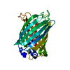
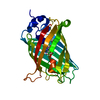
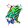
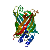
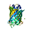
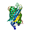
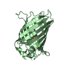
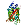
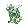
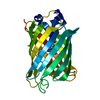
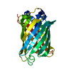
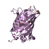
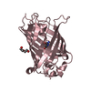
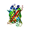
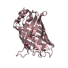
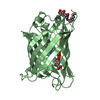
Green Fluorescent Protein / Green fluorescent protein / Green fluorescent protein, GFP / Green fluorescent protein-related / Green fluorescent protein / Green fluorescent protein / Beta Barrel / Mainly Beta
Similarity search - Domain/homology
D-MALATE / Green fluorescent protein
Similarity search - Component
Biological speciesAequorea victoria (jellyfish)
MethodX-RAY DIFFRACTION / MOLECULAR REPLACEMENT / Resolution: 2.49 Å
AuthorsRemington, S.J. / Trujillo, K.
Funding support United States, Israel, 2items
OrganizationGrant numberCountry
National Science Foundation (NSF, United States)MCB-1021374 United States
Binational Science FoundationBSF-2010082 Israel
CitationJournal: To Be Published
Title: Ultrafast Dynamics and Mechanisms of Proton Transfer in the GFP S205G Mutant
Authors: Wineman-Fisher, V. / Simkovitch, R. / Huppert, D. / Trujillo, K. / Remington, S.J. / Miller, Y.
History
DepositionFeb 2, 2016Deposition site: RCSB / Processing site: RCSB
Revision 1.0Feb 17, 2016Provider: repository / Type: Initial release
Revision 1.1Sep 27, 2017Group: Author supporting evidence / Derived calculations / Category: pdbx_audit_support / pdbx_struct_oper_list
Item: _pdbx_audit_support.funding_organization / _pdbx_struct_oper_list.symmetry_operation
Revision 1.2Nov 27, 2019Group: Author supporting evidence / Category: pdbx_audit_support / Item: _pdbx_audit_support.funding_organization
Revision 1.3Sep 27, 2023Group: Data collection / Database references / Refinement description
Category: chem_comp_atom / chem_comp_bond ...chem_comp_atom / chem_comp_bond / database_2 / pdbx_initial_refinement_model
Item: _database_2.pdbx_DOI / _database_2.pdbx_database_accession
Revision 2.0Nov 15, 2023Group: Atomic model / Data collection / Derived calculations
Category: atom_site / chem_comp_atom ...atom_site / chem_comp_atom / chem_comp_bond / pdbx_validate_torsion / struct_conn
Item: _atom_site.auth_atom_id / _atom_site.label_atom_id ..._atom_site.auth_atom_id / _atom_site.label_atom_id / _chem_comp_atom.atom_id / _chem_comp_bond.atom_id_1 / _chem_comp_bond.atom_id_2 / _struct_conn.pdbx_leaving_atom_flag / _struct_conn.ptnr1_label_atom_id / _struct_conn.ptnr2_label_atom_id

-
Structure visualization

Structure viewerMolecule:
MolmilJmol/JSmol

Downloads & links

-
Assembly

Deposited unit
A: Green fluorescent protein
B: Green fluorescent protein
hetero molecules


Theoretical massNumber of molelcules
Total (without water)53,7565
Polymers52,9922
Non-polymers7653
Water2,000111
1
A: Green fluorescent protein
hetero molecules


Theoretical massNumber of molelcules
Total (without water)26,6302
Polymers26,4961
Non-polymers1341
Water181
TypeNameSymmetry operationNumber
identity operation1_555x,y,z1
MethodPISA
2
B: Green fluorescent protein
hetero molecules


Theoretical massNumber of molelcules
Total (without water)27,1263
Polymers26,4961
Non-polymers6312
Water181
TypeNameSymmetry operationNumber
identity operation1_555x,y,z1
MethodPISA
Unit cell
Length a, b, c (Å)66.031, 96.284, 151.591
Angle α, β, γ (deg.)90.00, 90.00, 90.00
Int Tables number20
Space group name H-MC2221

-
Components

#1: Protein Green fluorescent protein /


Mass: 26495.816 Da / Num. of mol.: 2 / Fragment: UNP residues 1-235
Source method: isolated from a genetically manipulated source
Source: (gene. exp.) Aequorea victoria (jellyfish) / Gene: GFP / Production host: Escherichia coli (E. coli) / References: UniProt: P42212
#2: Chemical ChemComp-MLT / D-MALATE / (2R)-2-HYDROXYBUTANEDIOIC ACID / 2-HYDROXY-SUCCINIC ACID / Malic acid


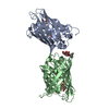
Mass: 134.087 Da / Num. of mol.: 2
Source method: isolated from a genetically manipulated source
Formula: C4H6O5
#3: Chemical ChemComp-UMQ / UNDECYL-MALTOSIDE / UNDECYL-BETA-D-MALTOPYRANOSIDE


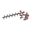
Mass: 496.589 Da / Num. of mol.: 1 / Source method: obtained synthetically / Formula: C23H44O11 / Comment: detergent*YM
#4: Water ChemComp-HOH / water / Water


Mass: 18.015 Da / Num. of mol.: 111 / Source method: isolated from a natural source / Formula: H2O

-
Experimental details

-
Experiment

ExperimentMethod: X-RAY DIFFRACTION / Number of used crystals: 1

-
Sample preparation

CrystalDensity Matthews: 2.31 Å3/Da / Density % sol: 46.75 %
Crystal growTemperature: 295 K / Method: vapor diffusion, hanging drop / pH: 7.9
Details: 1.0 M malate, pH 8.0, Tris, 0.3 M NaCl, 1% n-nonyl-beta-D maltoside

-
Data collection

DiffractionMean temperature: 100 K
Diffraction sourceSource: ROTATING ANODE / Type: RIGAKU RU300 / Wavelength: 1.54 Å
DetectorType: RIGAKU RAXIS IV / Detector: IMAGE PLATE / Date: Feb 24, 2010
RadiationMonochromator: Ni filter / Protocol: SINGLE WAVELENGTH / Monochromatic (M) / Laue (L): M / Scattering type: x-ray
Radiation wavelengthWavelength: 1.54 Å / Relative weight: 1
ReflectionResolution: 2.49→75 Å / Num. obs: 13474 / % possible obs: 96.9 % / Redundancy: 2.6 % / Rmerge(I) obs: 0.1 / Net I/σ(I): 13.8
Reflection shellResolution: 2.49→2.54 Å / Redundancy: 1.3 % / Rmerge(I) obs: 0.43 / Mean I/σ(I) obs: 1.3 / % possible all: 12.4

-
Processing

Software
NameVersionClassification
REFMAC5.8.0073refinement
HKL-2000data reduction
HKL-2000data scaling
EPMRphasing
RefinementMethod to determine structure: MOLECULAR REPLACEMENT
Starting model: 1EMB
Resolution: 2.49→10 Å / Cor.coef. Fo:Fc: 0.951 / Cor.coef. Fo:Fc free: 0.858 / SU B: 14.872 / SU ML: 0.317 / Cross valid method: THROUGHOUT / ESU R Free: 0.42 / Stereochemistry target values: MAXIMUM LIKELIHOOD / Details: HYDROGENS HAVE BEEN ADDED IN THE RIDING POSITIONS
RfactorNum. reflection% reflectionSelection details
Rfree0.27598 644 4.9 %RANDOM
Rwork0.175 ---
obs0.17993 12468 75.71 %-
Solvent computationIon probe radii: 0.8 Å / Shrinkage radii: 0.8 Å / VDW probe radii: 1.2 Å / Solvent model: MASK
Displacement parametersBiso mean: 38.05 Å2
Baniso -1Baniso -2Baniso -3
1--2.15 Å20 Å2-0 Å2
2--3.29 Å20 Å2
3----1.14 Å2
Refinement stepCycle: LAST / Resolution: 2.49→10 Å
ProteinNucleic acidLigandSolventTotal
Num. atoms3586 0 50 111 3747
Refine LS restraints
Refine-IDTypeDev idealDev ideal targetNumber
X-RAY DIFFRACTIONr_bond_refined_d0.0140.0193726
X-RAY DIFFRACTIONr_bond_other_d0.0040.023369
X-RAY DIFFRACTIONr_angle_refined_deg1.8531.9655054
X-RAY DIFFRACTIONr_angle_other_deg1.0033.0057746
X-RAY DIFFRACTIONr_dihedral_angle_1_deg8.6515454
X-RAY DIFFRACTIONr_dihedral_angle_2_deg33.27124.689177
X-RAY DIFFRACTIONr_dihedral_angle_3_deg18.49815569
X-RAY DIFFRACTIONr_dihedral_angle_4_deg11.8131514
X-RAY DIFFRACTIONr_chiral_restr0.0880.2549
X-RAY DIFFRACTIONr_gen_planes_refined0.0070.0214268
X-RAY DIFFRACTIONr_gen_planes_other0.0010.02878
X-RAY DIFFRACTIONr_nbd_refined
X-RAY DIFFRACTIONr_nbd_other
X-RAY DIFFRACTIONr_nbtor_refined
X-RAY DIFFRACTIONr_nbtor_other
X-RAY DIFFRACTIONr_xyhbond_nbd_refined
X-RAY DIFFRACTIONr_xyhbond_nbd_other
X-RAY DIFFRACTIONr_metal_ion_refined
X-RAY DIFFRACTIONr_metal_ion_other
X-RAY DIFFRACTIONr_symmetry_vdw_refined
X-RAY DIFFRACTIONr_symmetry_vdw_other
X-RAY DIFFRACTIONr_symmetry_hbond_refined
X-RAY DIFFRACTIONr_symmetry_hbond_other
X-RAY DIFFRACTIONr_symmetry_metal_ion_refined
X-RAY DIFFRACTIONr_symmetry_metal_ion_other
X-RAY DIFFRACTIONr_mcbond_it2.3813.8471827
X-RAY DIFFRACTIONr_mcbond_other2.3783.8481826
X-RAY DIFFRACTIONr_mcangle_it3.7735.7612276
X-RAY DIFFRACTIONr_mcangle_other3.7725.7622277
X-RAY DIFFRACTIONr_scbond_it2.5473.9831899
X-RAY DIFFRACTIONr_scbond_other2.5463.9811896
X-RAY DIFFRACTIONr_scangle_it
X-RAY DIFFRACTIONr_scangle_other4.0275.8952777
X-RAY DIFFRACTIONr_long_range_B_refined6.69630.5524059
X-RAY DIFFRACTIONr_long_range_B_other6.69730.5534045
X-RAY DIFFRACTIONr_rigid_bond_restr
X-RAY DIFFRACTIONr_sphericity_free
X-RAY DIFFRACTIONr_sphericity_bonded
LS refinement shellResolution: 2.49→2.551 Å / Total num. of bins used: 20
RfactorNum. reflection% reflection
Rfree0.302 14 -
Rwork0.309 171 -
obs--15.42 %

+
About Yorodumi

-
News

-
Feb 9, 2022. New format data for meta-information of EMDB entries

New format data for meta-information of EMDB entries

  • Version 3 of the EMDB header file is now the official format.
  • The previous official version 1.9 will be removed from the archive.

Related info.:EMDB header

External links:wwPDB to switch to version 3 of the EMDB data model

-
Aug 12, 2020. Covid-19 info

Covid-19 info

URL: https://pdbj.org/emnavi/covid19.php

New page: Covid-19 featured information page in EM Navigator.

Related info.:Covid-19 info / Mar 5, 2020. Novel coronavirus structure data

+
Mar 5, 2020. Novel coronavirus structure data

Novel coronavirus structure data

Related info.:Yorodumi Speices / Aug 12, 2020. Covid-19 info

External links:COVID-19 featured content - PDBj / Molecule of the Month (242):Coronavirus Proteases

+
Jan 31, 2019. EMDB accession codes are about to change! (news from PDBe EMDB page)

EMDB accession codes are about to change! (news from PDBe EMDB page)

  • The allocation of 4 digits for EMDB accession codes will soon come to an end. Whilst these codes will remain in use, new EMDB accession codes will include an additional digit and will expand incrementally as the available range of codes is exhausted. The current 4-digit format prefixed with “EMD-” (i.e. EMD-XXXX) will advance to a 5-digit format (i.e. EMD-XXXXX), and so on. It is currently estimated that the 4-digit codes will be depleted around Spring 2019, at which point the 5-digit format will come into force.
  • The EM Navigator/Yorodumi systems omit the EMD- prefix.

Related info.:Q: What is EMD? / ID/Accession-code notation in Yorodumi/EM Navigator

External links:EMDB Accession Codes are Changing Soon! / Contact to PDBj

+
Jul 12, 2017. Major update of PDB

Major update of PDB

  • wwPDB released updated PDB data conforming to the new PDBx/mmCIF dictionary.
  • This is a major update changing the version number from 4 to 5, and with Remediation, in which all the entries are updated.
  • In this update, many items about electron microscopy experimental information are reorganized (e.g. em_software).
  • Now, EM Navigator and Yorodumi are based on the updated data.

External links:wwPDB Remediation / Enriched Model Files Conforming to OneDep Data Standards Now Available in the PDB FTP Archive

-
Yorodumi

Thousand views of thousand structures

  • Yorodumi is a browser for structure data from EMDB, PDB, SASBDB, etc.
  • This page is also the successor to EM Navigator detail page, and also detail information page/front-end page for Omokage search.
  • The word "yorodu" (or yorozu) is an old Japanese word meaning "ten thousand". "mi" (miru) is to see.

Related info.:EMDB / PDB / SASBDB / Comparison of 3 databanks / Yorodumi Search / Aug 31, 2016. New EM Navigator & Yorodumi / Yorodumi Papers / Jmol/JSmol / Function and homology information / Changes in new EM Navigator and Yorodumi

Read more