 Movie
Movie Controller

[English] 日本語
 Yorodumi
- PDB-4xl2: Crystal structure of oxidized form of thiolase from Clostridium a... -
+ Open data

- Basic information
| Entry | Database: PDB / ID: 4xl2 | ||||||
|---|---|---|---|---|---|---|---|
| Title | Crystal structure of oxidized form of thiolase from Clostridium acetobutylicum | ||||||
 Components Components | Acetyl-CoA acetyltransferase | ||||||
 Keywords Keywords |  TRANSFERASE TRANSFERASE | ||||||
| Function / homology |  Function and homology informationacetyl-CoA C-acetyltransferase / acetyl-CoA C-acetyltransferase activity / cytoplasm Function and homology informationacetyl-CoA C-acetyltransferase / acetyl-CoA C-acetyltransferase activity / cytoplasmSimilarity search - Function | ||||||
| Biological species |  Clostridium acetobutylicum (bacteria) Clostridium acetobutylicum (bacteria) | ||||||
| Method | X-RAY DIFFRACTION / SYNCHROTRON / MOLECULAR REPLACEMENT / Resolution: 1.77 Å | ||||||
 Authors Authors | Kim, S. / Ha, S.C. / Ahn, J.W. / Kim, E.J. / Lim, J.H. / Kim, K.J. | ||||||
 Citation Citation | Journal: Nat Commun / Year: 2015 Title: Redox-switch regulatory mechanism of thiolase from Clostridium acetobutylicum Authors: Kim, S. / Jang, Y.S. / Ha, S.C. / Ahn, J.W. / Kim, E.J. / Hong Lim, J. / Cho, C. / Shin Ryu, Y. / Kuk Lee, S. / Lee, S.Y. / Kim, K.J. | ||||||
| History |
|
- Structure visualization
| Structure viewer | Molecule: MolmilJmol/JSmol |
|---|
- Downloads & links
-Download
| PDBx/mmCIF format | 4xl2.cif.gz | 167.2 KB | Display | PDBx/mmCIF format |
|---|---|---|---|---|
| PDB format | pdb4xl2.ent.gz | 131.2 KB | Display | PDB format |
| PDBx/mmJSON format | 4xl2.json.gz | Tree view | PDBx/mmJSON format | |
| Others |  Other downloads Other downloads |
-Validation report
| Arichive directory | https://data.pdbj.org/pub/pdb/validation_reports/xl/4xl2ftp://data.pdbj.org/pub/pdb/validation_reports/xl/4xl2 | HTTPS FTP |
|---|
-Related structure data
| Related structure data | 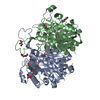 4wyrC  4wysC  4xl3C  4xl4C 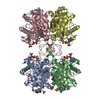 1dlvS C: citing same article ( S: Starting model for refinement |
|---|---|
| Similar structure data |


-Links
 PDBj
PDBj

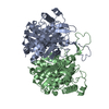
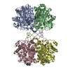
- Assembly
| Deposited unit | 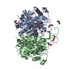
| ||||||||
|---|---|---|---|---|---|---|---|---|---|
| 1 | 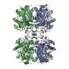
| ||||||||
| 2 |
| ||||||||
| Unit cell |
| ||||||||
| Details | biological unit is the same as asym. |
-Components
| #1: Protein | Mass: 42361.559 Da / Num. of mol.: 2 Source method: isolated from a genetically manipulated source Source: (gene. exp.) Clostridium acetobutylicum (strain ATCC 824 / DSM 792 / JCM 1419 / LMG 5710 / VKM B-1787) (bacteria)Strain: ATCC 824 / DSM 792 / JCM 1419 / LMG 5710 / VKM B-1787 Gene: thlA, thl, CA_C2873 / Plasmid: pET30a / Production host: Escherichia coli (E. coli) / Strain (production host): B834 / References: UniProt: P45359, acetyl-CoA C-acetyltransferase#2: Chemical | Acetate  Mass: 59.044 Da / Num. of mol.: 2 / Source method: obtained synthetically / Formula: C2H3O2 Mass: 59.044 Da / Num. of mol.: 2 / Source method: obtained synthetically / Formula: C2H3O2#3: Chemical | ChemComp-PEG / Diethylene glycol  Mass: 106.120 Da / Num. of mol.: 8 / Source method: obtained synthetically / Formula: C4H10O3 Mass: 106.120 Da / Num. of mol.: 8 / Source method: obtained synthetically / Formula: C4H10O3#4: Chemical | Glycerol  Mass: 92.094 Da / Num. of mol.: 2 / Source method: obtained synthetically / Formula: C3H8O3 Mass: 92.094 Da / Num. of mol.: 2 / Source method: obtained synthetically / Formula: C3H8O3#5: Water | ChemComp-HOH / | Water Mass: 18.015 Da / Num. of mol.: 488 / Source method: isolated from a natural source / Formula: H2O Mass: 18.015 Da / Num. of mol.: 488 / Source method: isolated from a natural source / Formula: H2O |
|---|
-Experimental details
-Experiment
| Experiment | Method: X-RAY DIFFRACTION / Number of used crystals: 1 |
|---|
- Sample preparation
| Crystal | Density Matthews: 2.42 Å3/Da / Density % sol: 49.11 % |
|---|---|
| Crystal grow | Temperature: 295 K / Method: vapor diffusion, hanging drop / pH: 4.2 / Details: PEG 3350, K-citrate, NaCl |
-Data collection
| Diffraction | Mean temperature: 100 K |
|---|---|
| Diffraction source | Source: SYNCHROTRON / Site: PAL/PLS  / Beamline: 7A (6B, 6C1) / Wavelength: 1.23985 Å / Beamline: 7A (6B, 6C1) / Wavelength: 1.23985 Å |
| Detector | Type: ADSC QUANTUM 210 / Detector: CCD / Date: Oct 17, 2008 |
| Radiation | Protocol: SINGLE WAVELENGTH / Monochromatic (M) / Laue (L): M / Scattering type: x-ray |
| Radiation wavelength | Wavelength: 1.23985 Å / Relative weight: 1 |
| Reflection | Resolution: 1.77→50 Å / Num. obs: 79061 / % possible obs: 98.1 % / Redundancy: 5.3 % / Rmerge(I) obs: 0.07 / Net I/σ(I): 33.7 |
| Reflection shell | Resolution: 1.77→1.83 Å / Redundancy: 4.1 % / Rmerge(I) obs: 0.299 / Mean I/σ(I) obs: 2.8 / % possible all: 87.9 |
- Processing
| Software |
| ||||||||||||||||||||||||||||||||||||||||||||||||||||||||||||||||||||||||||||||||||||||||||||||||||||||||||||||||||||||||||||||||||||||||||||||||||||||||||||||||||||||||||||||||||||||
|---|---|---|---|---|---|---|---|---|---|---|---|---|---|---|---|---|---|---|---|---|---|---|---|---|---|---|---|---|---|---|---|---|---|---|---|---|---|---|---|---|---|---|---|---|---|---|---|---|---|---|---|---|---|---|---|---|---|---|---|---|---|---|---|---|---|---|---|---|---|---|---|---|---|---|---|---|---|---|---|---|---|---|---|---|---|---|---|---|---|---|---|---|---|---|---|---|---|---|---|---|---|---|---|---|---|---|---|---|---|---|---|---|---|---|---|---|---|---|---|---|---|---|---|---|---|---|---|---|---|---|---|---|---|---|---|---|---|---|---|---|---|---|---|---|---|---|---|---|---|---|---|---|---|---|---|---|---|---|---|---|---|---|---|---|---|---|---|---|---|---|---|---|---|---|---|---|---|---|---|---|---|---|---|
| Refinement | Method to determine structure: MOLECULAR REPLACEMENT Starting model: 1DLV Resolution: 1.77→50 Å / Cor.coef. Fo:Fc: 0.972 / Cor.coef. Fo:Fc free: 0.96 / SU B: 2.564 / SU ML: 0.078 / Cross valid method: THROUGHOUT / ESU R: 0.101 / ESU R Free: 0.101 / Stereochemistry target values: MAXIMUM LIKELIHOOD / Details: HYDROGENS HAVE BEEN ADDED IN THE RIDING POSITIONS
| ||||||||||||||||||||||||||||||||||||||||||||||||||||||||||||||||||||||||||||||||||||||||||||||||||||||||||||||||||||||||||||||||||||||||||||||||||||||||||||||||||||||||||||||||||||||
| Solvent computation | Ion probe radii: 0.8 Å / Shrinkage radii: 0.8 Å / VDW probe radii: 1.2 Å / Solvent model: MASK | ||||||||||||||||||||||||||||||||||||||||||||||||||||||||||||||||||||||||||||||||||||||||||||||||||||||||||||||||||||||||||||||||||||||||||||||||||||||||||||||||||||||||||||||||||||||
| Displacement parameters | Biso mean: 32.174 Å2
| ||||||||||||||||||||||||||||||||||||||||||||||||||||||||||||||||||||||||||||||||||||||||||||||||||||||||||||||||||||||||||||||||||||||||||||||||||||||||||||||||||||||||||||||||||||||
| Refinement step | Cycle: LAST / Resolution: 1.77→50 Å
| ||||||||||||||||||||||||||||||||||||||||||||||||||||||||||||||||||||||||||||||||||||||||||||||||||||||||||||||||||||||||||||||||||||||||||||||||||||||||||||||||||||||||||||||||||||||
| Refine LS restraints |
|