 Movie
Movie Controller


+ Open data

- Basic information
| Entry | Database: PDB / ID: 4xd9 | ||||||
|---|---|---|---|---|---|---|---|
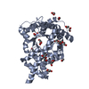
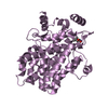
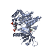
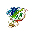
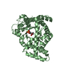
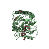
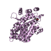
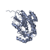
| Title | Structure of Rpf2-Rrs1 complex involved in ribosome biogenesis | ||||||
 Components Components |
| ||||||
 Keywords Keywords |  TRANSLATION / complex TRANSLATION / complex | ||||||
| Function / homology |  Function and homology information Function and homology informationpreribosome, large subunit precursor / endonucleolytic cleavage in ITS1 to separate SSU-rRNA from 5.8S rRNA and LSU-rRNA from tricistronic rRNA transcript (SSU-rRNA, 5.8S rRNA, LSU-rRNA) / maturation of LSU-rRNA from tricistronic rRNA transcript (SSU-rRNA, 5.8S rRNA, LSU-rRNA) / ribosomal large subunit biogenesis / ribosome assembly / ribosomal large subunit assembly / single-stranded RNA binding / rRNA binding / nucleolusSimilarity search - Function | ||||||
| Biological species |  Emericella nidulans (mold) Emericella nidulans (mold) | ||||||
| Method | X-RAY DIFFRACTION / SYNCHROTRON / Resolution: 2.35 Å | ||||||
 Authors Authors | Asano, N. / Kato, K. / Yao, M. | ||||||
| Funding support |  Japan, 1items Japan, 1items
| ||||||
 Citation Citation | Journal: Nucleic Acids Res. / Year: 2015 Title: Structural and functional analysis of the Rpf2-Rrs1 complex in ribosome biogenesis. Authors: Asano, N. / Kato, K. / Nakamura, A. / Komoda, K. / Tanaka, I. / Yao, M. | ||||||
| History |
|
- Structure visualization
| Structure viewer | Molecule: MolmilJmol/JSmol |
|---|
- Downloads & links
-Download
| PDBx/mmCIF format | 4xd9.cif.gz | 147.5 KB | Display | PDBx/mmCIF format |
|---|---|---|---|---|
| PDB format | pdb4xd9.ent.gz | 111.1 KB | Display | PDB format |
| PDBx/mmJSON format | 4xd9.json.gz | Tree view | PDBx/mmJSON format | |
| Others |  Other downloads Other downloads |
-Validation report
| Arichive directory | https://data.pdbj.org/pub/pdb/validation_reports/xd/4xd9ftp://data.pdbj.org/pub/pdb/validation_reports/xd/4xd9 | HTTPS FTP |
|---|
-Related structure data
| Similar structure data |
|---|


-Links
 PDBj
PDBj- Assembly
| Deposited unit | 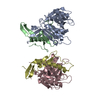
| ||||||||
|---|---|---|---|---|---|---|---|---|---|
| 1 | 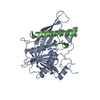
| ||||||||
| 2 | 
| ||||||||
| Unit cell |
|
-Components
| #1: Protein | / Uncharacterized protein Mass: 36407.578 Da / Num. of mol.: 2 / Fragment: UNP RESIDUES 1-302 Source method: isolated from a genetically manipulated source Source: (gene. exp.) Emericella nidulans (strain FGSC A4 / ATCC 38163 / CBS 112.46 / NRRL 194 / M139) (mold)Strain: FGSC A4 / ATCC 38163 / CBS 112.46 / NRRL 194 / M139 / Gene: AN3745.2, ANIA_03745 / Production host:  Escherichia coli (E. coli) / References: UniProt: C8VMF9 Escherichia coli (E. coli) / References: UniProt: C8VMF9#2: Protein | Mass: 25003.686 Da / Num. of mol.: 2 Source method: isolated from a genetically manipulated source Source: (gene. exp.) Emericella nidulans (strain FGSC A4 / ATCC 38163 / CBS 112.46 / NRRL 194 / M139) (mold)Strain: FGSC A4 / ATCC 38163 / CBS 112.46 / NRRL 194 / M139 / Gene: ANIA_10200 / Production host: Escherichia coli (E. coli) / References: UniProt: Q5B6T5#3: Water | ChemComp-HOH / | Water Mass: 18.015 Da / Num. of mol.: 163 / Source method: isolated from a natural source / Formula: H2O Mass: 18.015 Da / Num. of mol.: 163 / Source method: isolated from a natural source / Formula: H2O |
|---|
-Experimental details
-Experiment
| Experiment | Method: X-RAY DIFFRACTION |
|---|
- Sample preparation
| Crystal | Density Matthews: 1.82 Å3/Da / Density % sol: 32.54 % |
|---|---|
| Crystal grow | Temperature: 293 K / Method: vapor diffusion, sitting drop / Details: PEG 6000, HEPES |
-Data collection
| Diffraction | Mean temperature: 100 K |
|---|---|
| Diffraction source | Source: SYNCHROTRON / Site: SPring-8 / Beamline: BL41XU / Wavelength: 1 Å |
| Detector | Type: RAYONIX MX225HE / Detector: CCD / Date: Jul 4, 2012 |
| Radiation | Protocol: SINGLE WAVELENGTH / Monochromatic (M) / Laue (L): M / Scattering type: x-ray |
| Radiation wavelength | Wavelength: 1 Å / Relative weight: 1 |
| Reflection | Resolution: 2.35→50 Å / Num. obs: 38131 / % possible obs: 99.4 % / Redundancy: 7.2 % / Net I/σ(I): 14.51 |
- Processing
| Software |
| |||||||||||||||||||||||||||||||||||||||||||||||||||||||||||||||||||||||||||||||||||||||||||||||||||||||||
|---|---|---|---|---|---|---|---|---|---|---|---|---|---|---|---|---|---|---|---|---|---|---|---|---|---|---|---|---|---|---|---|---|---|---|---|---|---|---|---|---|---|---|---|---|---|---|---|---|---|---|---|---|---|---|---|---|---|---|---|---|---|---|---|---|---|---|---|---|---|---|---|---|---|---|---|---|---|---|---|---|---|---|---|---|---|---|---|---|---|---|---|---|---|---|---|---|---|---|---|---|---|---|---|---|---|---|
| Refinement | Resolution: 2.35→42.098 Å / SU ML: 0.3 / Cross valid method: FREE R-VALUE / σ(F): 1.35 / Phase error: 23.8 / Stereochemistry target values: ML
| |||||||||||||||||||||||||||||||||||||||||||||||||||||||||||||||||||||||||||||||||||||||||||||||||||||||||
| Solvent computation | Shrinkage radii: 0.9 Å / VDW probe radii: 1.11 Å / Solvent model: FLAT BULK SOLVENT MODEL | |||||||||||||||||||||||||||||||||||||||||||||||||||||||||||||||||||||||||||||||||||||||||||||||||||||||||
| Refinement step | Cycle: LAST / Resolution: 2.35→42.098 Å
| |||||||||||||||||||||||||||||||||||||||||||||||||||||||||||||||||||||||||||||||||||||||||||||||||||||||||
| Refine LS restraints |
| |||||||||||||||||||||||||||||||||||||||||||||||||||||||||||||||||||||||||||||||||||||||||||||||||||||||||
| LS refinement shell |
|
