 Movie
Movie Controller


+ Open data

- Basic information
| Entry | Database: PDB / ID: 3hrr | ||||||
|---|---|---|---|---|---|---|---|
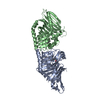
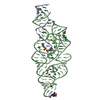
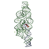
| Title | The Product Template Domain from PksA with Harris Compound Bound | ||||||
 Components Components | Aflatoxin biosynthesis polyketide synthase | ||||||
 Keywords Keywords |  TRANSCRIPTION / PksA / aflatoxin / norsolorinic acid / polyketide / polyketide synthase / PKS / iterative Type I PKS / Harris Compound / hot-dog fold / Acyltransferase / Multifunctional enzyme / Phosphopantetheine / Transferase TRANSCRIPTION / PksA / aflatoxin / norsolorinic acid / polyketide / polyketide synthase / PKS / iterative Type I PKS / Harris Compound / hot-dog fold / Acyltransferase / Multifunctional enzyme / Phosphopantetheine / Transferase | ||||||
| Function / homology |  Function and homology information Function and homology informationnoranthrone synthase / aflatoxin biosynthetic process / norsolorinate anthrone synthase activity / phosphopantetheine binding / identical protein bindingSimilarity search - Function | ||||||
| Biological species |  Aspergillus parasiticus (mold) Aspergillus parasiticus (mold) | ||||||
| Method | X-RAY DIFFRACTION / SYNCHROTRON / MOLECULAR REPLACEMENT / Resolution: 1.9 Å | ||||||
 Authors Authors | Korman, T.P. / Tsai, S.C. | ||||||
 Citation Citation | Journal: Nature / Year: 2009 Title: Structural basis for biosynthetic programming of fungal aromatic polyketide cyclization. Authors: Crawford, J.M. / Korman, T.P. / Labonte, J.W. / Vagstad, A.L. / Hill, E.A. / Kamari-Bidkorpeh, O. / Tsai, S.C. / Townsend, C.A. | ||||||
| History |
|
- Structure visualization
| Structure viewer | Molecule: MolmilJmol/JSmol |
|---|
- Downloads & links
-Download
| PDBx/mmCIF format | 3hrr.cif.gz | 145.1 KB | Display | PDBx/mmCIF format |
|---|---|---|---|---|
| PDB format | pdb3hrr.ent.gz | 112 KB | Display | PDB format |
| PDBx/mmJSON format | 3hrr.json.gz | Tree view | PDBx/mmJSON format | |
| Others |  Other downloads Other downloads |
-Validation report
| Arichive directory | https://data.pdbj.org/pub/pdb/validation_reports/hr/3hrrftp://data.pdbj.org/pub/pdb/validation_reports/hr/3hrr | HTTPS FTP |
|---|
-Related structure data
| Related structure data |  3hrqSC S: Starting model for refinement C: citing same article ( |
|---|---|
| Similar structure data |


-Links
 PDBj
PDBj


- Assembly
| Deposited unit | 
| ||||||||
|---|---|---|---|---|---|---|---|---|---|
| 1 |
| ||||||||
| 2 | 
| ||||||||
| 3 | 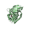
| ||||||||
| Unit cell |
|
-Components
| #1: Protein | Mass: 39243.543 Da / Num. of mol.: 2 / Fragment: UNP residues 1305-1660 Source method: isolated from a genetically manipulated source Source: (gene. exp.) Aspergillus parasiticus (mold) / Gene: PksA, pksL1 / Production host:  Escherichia coli (E. coli) / Strain (production host): BL21(DE3) / References: UniProt: Q12053 Escherichia coli (E. coli) / Strain (production host): BL21(DE3) / References: UniProt: Q12053#2: Chemical |   Mass: 288.295 Da / Num. of mol.: 2 / Source method: obtained synthetically / Formula: C16H16O5 Mass: 288.295 Da / Num. of mol.: 2 / Source method: obtained synthetically / Formula: C16H16O5#3: Water | ChemComp-HOH / | Water Mass: 18.015 Da / Num. of mol.: 392 / Source method: isolated from a natural source / Formula: H2O Mass: 18.015 Da / Num. of mol.: 392 / Source method: isolated from a natural source / Formula: H2O |
|---|
-Experimental details
-Experiment
| Experiment | Method: X-RAY DIFFRACTION / Number of used crystals: 1 |
|---|
- Sample preparation
| Crystal | Density Matthews: 2.32 Å3/Da / Density % sol: 47.07 % |
|---|---|
| Crystal grow | Temperature: 298 K / Method: vapor diffusion / pH: 7.5 Details: 0.22 M Ammonium Acetate, 20% PEG3350, pH 7.5, VAPOR DIFFUSION, temperature 298K |
-Data collection
| Diffraction | Mean temperature: 100 K | ||||||||||||||||||||||||||||||||||||||||||||||||||||||||||||||||||
|---|---|---|---|---|---|---|---|---|---|---|---|---|---|---|---|---|---|---|---|---|---|---|---|---|---|---|---|---|---|---|---|---|---|---|---|---|---|---|---|---|---|---|---|---|---|---|---|---|---|---|---|---|---|---|---|---|---|---|---|---|---|---|---|---|---|---|---|
| Diffraction source | Source: SYNCHROTRON / Site: SSRL  / Beamline: BL9-1 / Wavelength: 1 Å / Beamline: BL9-1 / Wavelength: 1 Å | ||||||||||||||||||||||||||||||||||||||||||||||||||||||||||||||||||
| Detector | Type: ADSC QUANTUM 315 / Detector: CCD / Date: Jan 4, 2008 Details: Vertical focusing mirror; single crystal Si(311) bent monochromator (horizontal focusing) | ||||||||||||||||||||||||||||||||||||||||||||||||||||||||||||||||||
| Radiation | Monochromator: Side-scattering cuberoot I-beam bent single crystal; asymmetric cut 12.2 degs Protocol: SINGLE WAVELENGTH / Scattering type: x-ray | ||||||||||||||||||||||||||||||||||||||||||||||||||||||||||||||||||
| Radiation wavelength | Wavelength: 1 Å / Relative weight: 1 | ||||||||||||||||||||||||||||||||||||||||||||||||||||||||||||||||||
| Reflection | Resolution: 1.9→50 Å / Num. all: 58378 / Num. obs: 57678 / % possible obs: 98.8 % / Redundancy: 7 % / Rmerge(I) obs: 0.06 / Χ2: 0.987 / Net I/σ(I): 30.643 | ||||||||||||||||||||||||||||||||||||||||||||||||||||||||||||||||||
| Reflection shell |
|
- Processing
| Software |
| ||||||||||||||||||||||||||||||||||||||||||||||||||||||||||||||||||||||||||||||||||||||||||
|---|---|---|---|---|---|---|---|---|---|---|---|---|---|---|---|---|---|---|---|---|---|---|---|---|---|---|---|---|---|---|---|---|---|---|---|---|---|---|---|---|---|---|---|---|---|---|---|---|---|---|---|---|---|---|---|---|---|---|---|---|---|---|---|---|---|---|---|---|---|---|---|---|---|---|---|---|---|---|---|---|---|---|---|---|---|---|---|---|---|---|---|
| Refinement | Method to determine structure: MOLECULAR REPLACEMENT Starting model: PDB entry 3HRQ Resolution: 1.9→44.4 Å / Cor.coef. Fo:Fc: 0.949 / Cor.coef. Fo:Fc free: 0.934 / Occupancy max: 1 / Occupancy min: 1 / SU B: 2.867 / SU ML: 0.086 / Cross valid method: THROUGHOUT / σ(F): 0 / ESU R: 0.149 / ESU R Free: 0.136 / Stereochemistry target values: MAXIMUM LIKELIHOOD / Details: HYDROGENS HAVE BEEN ADDED IN THE RIDING POSITIONS
| ||||||||||||||||||||||||||||||||||||||||||||||||||||||||||||||||||||||||||||||||||||||||||
| Solvent computation | Ion probe radii: 0.8 Å / Shrinkage radii: 0.8 Å / VDW probe radii: 1.2 Å / Solvent model: MASK | ||||||||||||||||||||||||||||||||||||||||||||||||||||||||||||||||||||||||||||||||||||||||||
| Displacement parameters | Biso max: 70.51 Å2 / Biso mean: 25.291 Å2 / Biso min: 8.81 Å2
| ||||||||||||||||||||||||||||||||||||||||||||||||||||||||||||||||||||||||||||||||||||||||||
| Refinement step | Cycle: LAST / Resolution: 1.9→44.4 Å
| ||||||||||||||||||||||||||||||||||||||||||||||||||||||||||||||||||||||||||||||||||||||||||
| Refine LS restraints |
| ||||||||||||||||||||||||||||||||||||||||||||||||||||||||||||||||||||||||||||||||||||||||||
| LS refinement shell | Resolution: 1.9→1.949 Å / Total num. of bins used: 20
|
