- PDB-4xae: Structure of Feruloyl-CoA 6-hydroxylase (F6H) from Arabidopsis th... -
+
Open data
ID or keywords:
Loading...
-
Basic information
Entry
Database: PDB / ID: 4xae
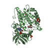
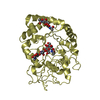
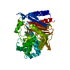
Title
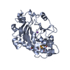
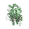
Structure of Feruloyl-CoA 6-hydroxylase (F6H) from Arabidopsis thaliana
Components
Feruloyl CoA ortho-hydroxylase 1
Keywords
OXIDOREDUCTASE / PROTEIN ENGINEERING / COUMARINS / 2-oxoglutarate dependent dioxygenase
Function / homology
Function and homology information
feruloyl-CoA 6-hydroxylase / hydrogen peroxide-mediated programmed cell death / response to iron ion starvation / coumarin biosynthetic process / phenylpropanoid biosynthetic process / dioxygenase activity / metal ion binding Similarity search - Function
Mass: 42306.938 Da / Num. of mol.: 2 Source method: isolated from a genetically manipulated source Source: (gene. exp.) Arabidopsis thaliana (thale cress) / Gene: F6'H1, At3g13610, K20M4.5 / Production host: Escherichia coli (E. coli) References: UniProt: Q9LHN8, Oxidoreductases; Acting on paired donors, with incorporation or reduction of molecular oxygen; With 2-oxoglutarate as one donor, and incorporation of one atom of oxygen into each donor
Mass: 18.015 Da / Num. of mol.: 28 / Source method: isolated from a natural source / Formula: H2O
-
Experimental details
-
Experiment
Experiment
Method: X-RAY DIFFRACTION
-
Sample preparation
Crystal
Density Matthews: 2.28 Å3/Da / Density % sol: 46.13 %
Crystal grow
Temperature: 291 K / Method: vapor diffusion, sitting drop / pH: 6 Details: Sitting drop vapor diffusion at 291K using 2 microliter drops containing equal volumes of protein concentrate and a precipitant cocktail containing 20% (w/v) PEG-8000, 0.1 M MES, 0.3 M Ca(OAc)2, pH 6.0
In the structure databanks used in Yorodumi, some data are registered as the other names, "COVID-19 virus" and "2019-nCoV". Here are the details of the virus and the list of structure data.
Jan 31, 2019. EMDB accession codes are about to change! (news from PDBe EMDB page)
EMDB accession codes are about to change! (news from PDBe EMDB page)
The allocation of 4 digits for EMDB accession codes will soon come to an end. Whilst these codes will remain in use, new EMDB accession codes will include an additional digit and will expand incrementally as the available range of codes is exhausted. The current 4-digit format prefixed with “EMD-” (i.e. EMD-XXXX) will advance to a 5-digit format (i.e. EMD-XXXXX), and so on. It is currently estimated that the 4-digit codes will be depleted around Spring 2019, at which point the 5-digit format will come into force.
The EM Navigator/Yorodumi systems omit the EMD- prefix.
Related info.:Q: What is EMD? / ID/Accession-code notation in Yorodumi/EM Navigator
Yorodumi is a browser for structure data from EMDB, PDB, SASBDB, etc.
This page is also the successor to EM Navigator detail page, and also detail information page/front-end page for Omokage search.
The word "yorodu" (or yorozu) is an old Japanese word meaning "ten thousand". "mi" (miru) is to see.
Related info.:EMDB / PDB / SASBDB / Comparison of 3 databanks / Yorodumi Search / Aug 31, 2016. New EM Navigator & Yorodumi / Yorodumi Papers / Jmol/JSmol / Function and homology information / Changes in new EM Navigator and Yorodumi
 Movie
Movie Controller

 Yorodumi
 Open data
 Basic information
 Components
 Keywords
 OXIDOREDUCTASE /
  Function and homology information
 Authors
 Citation
 Structure visualization
 Downloads & links
 Other downloads
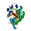


 PDBj
PDBj
 Assembly
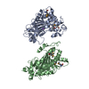


 Mass: 22.990 Da / Num. of mol.: 2 / Source method: obtained synthetically / Formula: Na
 Mass: 18.015 Da / Num. of mol.: 28 / Source method: isolated from a natural source / Formula: H2O
 Sample preparation
 / Beamline: 22-ID / Wavelength: 0.979 Å
 Processing