 Movie
Movie Controller

[English] 日本語
 Yorodumi
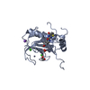
- PDB-4x2w: Crystal structure of the Murine Norovirus NS6 protease (inactive ... -
+ Open data

- Basic information
| Entry | Database: PDB / ID: 4x2w | ||||||
|---|---|---|---|---|---|---|---|
| Title | Crystal structure of the Murine Norovirus NS6 protease (inactive C139A mutant) with a C-terminal extension to include residues P1 prime - P2 prime of NS7 | ||||||
 Components Components | NS6 Protease | ||||||
 Keywords Keywords |  HYDROLASE / Murine norovirus Protease HYDROLASE / Murine norovirus Protease | ||||||
| Function / homology |  Function and homology informationcalicivirin / ribonucleoside triphosphate phosphatase activity / nucleoside-triphosphate phosphatase / RNA helicase activity / RNA-directed RNA polymerase / viral RNA genome replication / cysteine-type endopeptidase activity / RNA-dependent RNA polymerase activity / DNA-templated transcription / proteolysis ...calicivirin / ribonucleoside triphosphate phosphatase activity / nucleoside-triphosphate phosphatase / RNA helicase activity / RNA-directed RNA polymerase / viral RNA genome replication / cysteine-type endopeptidase activity / RNA-dependent RNA polymerase activity / DNA-templated transcription / proteolysis / RNA binding / ATP binding / metal ion binding Function and homology informationcalicivirin / ribonucleoside triphosphate phosphatase activity / nucleoside-triphosphate phosphatase / RNA helicase activity / RNA-directed RNA polymerase / viral RNA genome replication / cysteine-type endopeptidase activity / RNA-dependent RNA polymerase activity / DNA-templated transcription / proteolysis ...calicivirin / ribonucleoside triphosphate phosphatase activity / nucleoside-triphosphate phosphatase / RNA helicase activity / RNA-directed RNA polymerase / viral RNA genome replication / cysteine-type endopeptidase activity / RNA-dependent RNA polymerase activity / DNA-templated transcription / proteolysis / RNA binding / ATP binding / metal ion bindingSimilarity search - Function | ||||||
| Biological species |   Murine norovirus 1 Murine norovirus 1 | ||||||
| Method | X-RAY DIFFRACTION / SYNCHROTRON / MOLECULAR REPLACEMENT / Resolution: 2.7 Å | ||||||
 Authors Authors | Fernandes, H. / Leen, E.N. / Curry, S. | ||||||
 Citation Citation | Journal: Peerj / Year: 2015 Title: Structure determination of Murine Norovirus NS6 proteases with C-terminal extensions designed to probe protease-substrate interactions. Authors: Fernandes, H. / Leen, E.N. / Cromwell, H. / Pfeil, M.P. / Curry, S. #1: Journal: PLoS ONE / Year: 2012Title: Structure of a murine norovirus NS6 protease-product complex revealed by adventitious crystallisation. Authors: Leen, E.N. / Baeza, G. / Curry, S. | ||||||
| History |
|
- Structure visualization
| Structure viewer | Molecule: MolmilJmol/JSmol |
|---|
- Downloads & links
-Download
| PDBx/mmCIF format | 4x2w.cif.gz | 143.6 KB | Display | PDBx/mmCIF format |
|---|---|---|---|---|
| PDB format | pdb4x2w.ent.gz | 115.5 KB | Display | PDB format |
| PDBx/mmJSON format | 4x2w.json.gz | Tree view | PDBx/mmJSON format | |
| Others |  Other downloads Other downloads |
-Validation report
| Arichive directory | https://data.pdbj.org/pub/pdb/validation_reports/x2/4x2wftp://data.pdbj.org/pub/pdb/validation_reports/x2/4x2w | HTTPS FTP |
|---|
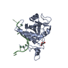
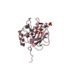
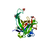
-Related structure data
| Related structure data |  4x2vC 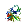 4x2xC  4x2yC 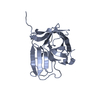 4ashS S: Starting model for refinement C: citing same article ( |
|---|---|
| Similar structure data |


-Links
 PDBj
PDBj
- Assembly
| Deposited unit | 
| ||||||||||||||||||
|---|---|---|---|---|---|---|---|---|---|---|---|---|---|---|---|---|---|---|---|
| 1 | 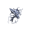
| ||||||||||||||||||
| 2 | 
| ||||||||||||||||||
| Unit cell |
| ||||||||||||||||||
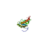
| Noncrystallographic symmetry (NCS) | NCS domain:
NCS domain segments: Component-ID: 0 / Ens-ID: 1 / Beg auth comp-ID: SER / Beg label comp-ID: SER / End auth comp-ID: GLU / End label comp-ID: GLU / Refine code: 0 / Auth seq-ID: 4 - 181 / Label seq-ID: 2 - 179
|
-Components
| #1: Protein | Mass: 18780.504 Da / Num. of mol.: 2 / Fragment: UNP residues 997-1175 / Mutation: C139A Source method: isolated from a genetically manipulated source Source: (gene. exp.) Murine norovirus 1 / Production host:  Escherichia coli BL21(DE3) (bacteria) / Variant (production host): CodonPlus / References: UniProt: Q80J95 Escherichia coli BL21(DE3) (bacteria) / Variant (production host): CodonPlus / References: UniProt: Q80J95 |
|---|
-Experimental details
-Experiment
| Experiment | Method: X-RAY DIFFRACTION / Number of used crystals: 1 |
|---|
- Sample preparation
| Crystal | Density Matthews: 2.93 Å3/Da / Density % sol: 57.95 % |
|---|---|
| Crystal grow | Temperature: 291.15 K / Method: vapor diffusion, sitting drop / pH: 7 Details: 15% (v/v) PEG 3350, 0.1 M glycine, 0.1 M Na-citrate pH 7.0 |
-Data collection
| Diffraction | Mean temperature: 100 K |
|---|---|
| Diffraction source | Source: SYNCHROTRON / Site: Diamond  / Beamline: I03 / Wavelength: 1 Å / Beamline: I03 / Wavelength: 1 Å |
| Detector | Type: DECTRIS PILATUS 6M-F / Detector: PIXEL / Date: Apr 21, 2013 |
| Radiation | Protocol: SINGLE WAVELENGTH / Monochromatic (M) / Laue (L): M / Scattering type: x-ray |
| Radiation wavelength | Wavelength: 1 Å / Relative weight: 1 |
| Reflection | Resolution: 2.7→117.85 Å / Num. obs: 8555 / % possible obs: 99.8 % / Redundancy: 5.7 % / Rmerge(I) obs: 0.085 / Net I/σ(I): 10.9 |
| Reflection shell | Resolution: 3.1→3.31 Å / Redundancy: 6 % / Rmerge(I) obs: 0.915 / Mean I/σ(I) obs: 2.1 / % possible all: 99.6 |
- Processing
| Software |
| |||||||||||||||||||||||||||||||||||||||||||||||||||||||||||||||||||||||||||
|---|---|---|---|---|---|---|---|---|---|---|---|---|---|---|---|---|---|---|---|---|---|---|---|---|---|---|---|---|---|---|---|---|---|---|---|---|---|---|---|---|---|---|---|---|---|---|---|---|---|---|---|---|---|---|---|---|---|---|---|---|---|---|---|---|---|---|---|---|---|---|---|---|---|---|---|---|
| Refinement | Method to determine structure: MOLECULAR REPLACEMENT Starting model: 4ASH Resolution: 2.7→117.85 Å / Cor.coef. Fo:Fc: 0.911 / Cor.coef. Fo:Fc free: 0.863 / SU B: 67.36 / SU ML: 0.534 / Cross valid method: FREE R-VALUE / σ(F): 0 / ESU R Free: 0.487 / Stereochemistry target values: MAXIMUM LIKELIHOOD / Details: HYDROGENS HAVE BEEN ADDED IN THE RIDING POSITIONS
| |||||||||||||||||||||||||||||||||||||||||||||||||||||||||||||||||||||||||||
| Solvent computation | Ion probe radii: 0.8 Å / Shrinkage radii: 0.8 Å / VDW probe radii: 1.2 Å / Solvent model: MASK | |||||||||||||||||||||||||||||||||||||||||||||||||||||||||||||||||||||||||||
| Displacement parameters | Biso max: 212.26 Å2 / Biso mean: 87.171 Å2 / Biso min: 42.52 Å2
| |||||||||||||||||||||||||||||||||||||||||||||||||||||||||||||||||||||||||||
| Refinement step | Cycle: final / Resolution: 2.7→117.85 Å
| |||||||||||||||||||||||||||||||||||||||||||||||||||||||||||||||||||||||||||
| Refine LS restraints |
| |||||||||||||||||||||||||||||||||||||||||||||||||||||||||||||||||||||||||||
| Refine LS restraints NCS | Ens-ID: 1 / Number: 8802 / Refine-ID: X-RAY DIFFRACTION / Type: interatomic distance / Rms dev position: 0.17 Å / Weight position: 0.05
| |||||||||||||||||||||||||||||||||||||||||||||||||||||||||||||||||||||||||||
| LS refinement shell | Resolution: 2.7→2.77 Å / Total num. of bins used: 20
| |||||||||||||||||||||||||||||||||||||||||||||||||||||||||||||||||||||||||||
| Refinement TLS params. | Method: refined / Refine-ID: X-RAY DIFFRACTION
| |||||||||||||||||||||||||||||||||||||||||||||||||||||||||||||||||||||||||||
| Refinement TLS group |
|
