- PDB-4x28: Crystal structure of the ChsE4-ChsE5 complex from Mycobacterium t... -
+
Open data
ID or keywords:
Loading...
-
Basic information
Entry
Database: PDB / ID: 4x28
Title
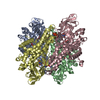
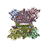
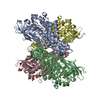
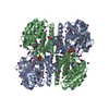
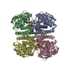
Crystal structure of the ChsE4-ChsE5 complex from Mycobacterium tuberculosis
Components
(Acyl-CoA dehydrogenase) x 2
Keywords
OXIDOREDUCTASE / Dehydrogenase
Function / homology
Function and homology information
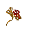
Oxidoreductases; Acting on the CH-CH group of donors; With unknown physiological acceptors / oxidoreductase activity, acting on the CH-CH group of donors / cholesterol catabolic process / : / flavin adenine dinucleotide binding Similarity search - Function
Protocol: SINGLE WAVELENGTH / Monochromatic (M) / Laue (L): M / Scattering type: x-ray
Radiation wavelength
Wavelength: 0.9792 Å / Relative weight: 1
Reflection
Resolution: 1.99→46.4 Å / Num. all: 739658 / Num. obs: 109767 / % possible obs: 99 % / Redundancy: 6.7 % / Rmerge(I) obs: 0.076 / Net I/σ(I): 19.9 / Phase calculation details: SAD
Reflection shell
Resolution: 1.99→2 Å / Redundancy: 6.6 % / Rmerge(I) obs: 0.624 / Mean I/σ(I) obs: 2.9 / % possible all: 99
-
Processing
Software
Name
Version
Classification
REFMAC
5.8.0073
refinement
XDS
datareduction
Aimless
datascaling
SHARP
phasing
Refinement
Method to determine structure: SAD / Resolution: 1.99→46.4 Å / Cor.coef. Fo:Fc: 0.974 / Cor.coef. Fo:Fc free: 0.962 / SU B: 3.844 / SU ML: 0.103 / Cross valid method: THROUGHOUT / ESU R: 0.146 / ESU R Free: 0.13 / Stereochemistry target values: MAXIMUM LIKELIHOOD / Details: HYDROGENS HAVE BEEN ADDED IN THE RIDING POSITIONS
Rfactor
Num. reflection
% reflection
Selection details
Rfree
0.18615
5476
5 %
RANDOM
Rwork
0.15079
-
-
-
obs
0.15261
-
99.9 %
-
Solvent computation
Ion probe radii: 0.8 Å / Shrinkage radii: 0.8 Å / VDW probe radii: 1.2 Å / Solvent model: MASK
 Movie
Movie Controller

 Yorodumi
 Open data
 Basic information
 Components
 ) x 2
) x 2  Keywords
 Function and homology information

 Authors
 United States, 2items
  Citation
 Structure visualization
 Downloads & links
 Other downloads
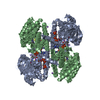


 PDBj
PDBj Assembly
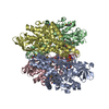

 Mass: 787.566 Da / Num. of mol.: 2 / Source method: obtained synthetically / Formula: C27H35N9O15P2
 Mass: 18.015 Da / Num. of mol.: 899 / Source method: isolated from a natural source / Formula: H2O
 Sample preparation
 Processing