 Movie
Movie Controller

[English] 日本語
 Yorodumi
- PDB-4x0m: Selection of fragments for kinase inhibitor design: decoration is key -
+ Open data

- Basic information
| Entry | Database: PDB / ID: 4x0m | ||||||
|---|---|---|---|---|---|---|---|
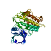
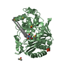
| Title | Selection of fragments for kinase inhibitor design: decoration is key | ||||||
 Components Components | TGF-beta receptor type-1 | ||||||
 Keywords Keywords |  TRANSFERASE / PROTEIN KINASE / INHIBITOR COMPLEX TRANSFERASE / PROTEIN KINASE / INHIBITOR COMPLEX | ||||||
| Function / homology |  Function and homology information Function and homology informationextracellular structure organization / epicardium morphogenesis / parathyroid gland development / transforming growth factor beta ligand-receptor complex / regulation of cardiac muscle cell proliferation / myofibroblast differentiation / positive regulation of tight junction disassembly / positive regulation of epithelial to mesenchymal transition involved in endocardial cushion formation / TGFBR2 Kinase Domain Mutants in Cancer / transforming growth factor beta receptor activity ...extracellular structure organization / epicardium morphogenesis / parathyroid gland development / transforming growth factor beta ligand-receptor complex / regulation of cardiac muscle cell proliferation / myofibroblast differentiation / positive regulation of tight junction disassembly / positive regulation of epithelial to mesenchymal transition involved in endocardial cushion formation / TGFBR2 Kinase Domain Mutants in Cancer / transforming growth factor beta receptor activity / positive regulation of mesenchymal stem cell proliferation / ventricular compact myocardium morphogenesis / SMAD2/3 Phosphorylation Motif Mutants in Cancer / TGFBR1 KD Mutants in Cancer / positive regulation of vasculature development / regulation of epithelial to mesenchymal transition / activin receptor activity, type I / cardiac epithelial to mesenchymal transition / transforming growth factor beta receptor activity, type I / activin receptor complex / mesenchymal cell differentiation / neuron fate commitment / germ cell migration / positive regulation of extracellular matrix assembly / type II transforming growth factor beta receptor binding / TGFBR1 LBD Mutants in Cancer / angiogenesis involved in coronary vascular morphogenesis / receptor protein serine/threonine kinase / transmembrane receptor protein serine/threonine kinase activity / pharyngeal system development / activin binding / coronary artery morphogenesis / activin receptor signaling pathway / filopodium assembly / ventricular trabecula myocardium morphogenesis / transforming growth factor beta binding / embryonic cranial skeleton morphogenesis / response to cholesterol / I-SMAD binding / negative regulation of chondrocyte differentiation / collagen fibril organization / endothelial cell proliferation / endothelial cell activation / skeletal system morphogenesis / lens development in camera-type eye / positive regulation of filopodium assembly / anterior/posterior pattern specification / artery morphogenesis / ventricular septum morphogenesis / roof of mouth development / TGF-beta receptor signaling activates SMADs / SMAD binding / negative regulation of endothelial cell proliferation / positive regulation of SMAD protein signal transduction / blastocyst development / regulation of protein ubiquitination / endothelial cell migration / bicellular tight junction / epithelial to mesenchymal transition / positive regulation of epithelial to mesenchymal transition / positive regulation of apoptotic signaling pathway / positive regulation of stress fiber assembly / cellular response to transforming growth factor beta stimulus / positive regulation of endothelial cell proliferation / Downregulation of TGF-beta receptor signaling / post-embryonic development / transforming growth factor beta receptor signaling pathway / negative regulation of cell migration / thymus development / TGF-beta receptor signaling in EMT (epithelial to mesenchymal transition) / kidney development / skeletal system development / cell motility / negative regulation of extrinsic apoptotic signaling pathway / wound healing / cellular response to growth factor stimulus / male gonad development / UCH proteinases / nervous system development / heart development / positive regulation of cell growth / regulation of gene expression / peptidyl-serine phosphorylation / in utero embryonic development / positive regulation of phosphatidylinositol 3-kinase/protein kinase B signal transduction / receptor complex / regulation of cell cycle / Ub-specific processing proteases / endosome / protein kinase activity / intracellular signal transduction / positive regulation of cell migration / membrane raft / protein serine/threonine kinase activity / apoptotic process / ubiquitin protein ligase binding / positive regulation of cell population proliferation / positive regulation of gene expression / regulation of DNA-templated transcription / positive regulation of DNA-templated transcriptionSimilarity search - Function | ||||||
| Biological species |  Homo sapiens (human) Homo sapiens (human) | ||||||
| Method | X-RAY DIFFRACTION / SYNCHROTRON / MOLECULAR REPLACEMENT / Resolution: 1.68 Å | ||||||
 Authors Authors | Czodrowski, P. / Hoelzemann, G. / Barnickel, G. / Greiner, H. / Musil, D. | ||||||
 Citation Citation | Journal: J.Med.Chem. / Year: 2015 Title: Selection of fragments for kinase inhibitor design: decoration is key. Authors: Czodrowski, P. / Holzemann, G. / Barnickel, G. / Greiner, H. / Musil, D. | ||||||
| History |
|
- Structure visualization
| Structure viewer | Molecule: MolmilJmol/JSmol |
|---|
- Downloads & links
-Download
| PDBx/mmCIF format | 4x0m.cif.gz | 136.5 KB | Display | PDBx/mmCIF format |
|---|---|---|---|---|
| PDB format | pdb4x0m.ent.gz | 105.1 KB | Display | PDB format |
| PDBx/mmJSON format | 4x0m.json.gz | Tree view | PDBx/mmJSON format | |
| Others |  Other downloads Other downloads |
-Validation report
| Arichive directory | https://data.pdbj.org/pub/pdb/validation_reports/x0/4x0mftp://data.pdbj.org/pub/pdb/validation_reports/x0/4x0m | HTTPS FTP |
|---|
-Related structure data
| Related structure data |  4x2fC 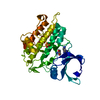 4x2gC  4x2jC  4x2kC 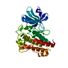 4x2nC 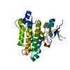 4x3jC 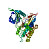 1vjyS S: Starting model for refinement C: citing same article ( |
|---|---|
| Similar structure data |


-Links
 PDBj
PDBj


- Assembly
| Deposited unit | 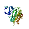
| ||||||||
|---|---|---|---|---|---|---|---|---|---|
| 1 |
| ||||||||
| Unit cell |
|
-Components
| #1: Protein | Mass: 34882.148 Da / Num. of mol.: 1 / Fragment: UNP residues 200-503 Source method: isolated from a genetically manipulated source Source: (gene. exp.) Homo sapiens (human) / Gene: TGFBR1, ALK5, SKR4 / Production host:   Spodoptera frugiperda (fall armyworm) Spodoptera frugiperda (fall armyworm)References: UniProt: P36897, receptor protein serine/threonine kinase |
|---|---|
| #2: Chemical | ChemComp-3WA /   Mass: 162.149 Da / Num. of mol.: 1 / Source method: obtained synthetically / Formula: C7H6N4O Mass: 162.149 Da / Num. of mol.: 1 / Source method: obtained synthetically / Formula: C7H6N4O |
| #3: Chemical | ChemComp-SO4 / Sulfate  Mass: 96.063 Da / Num. of mol.: 1 / Source method: obtained synthetically / Formula: SO4 Mass: 96.063 Da / Num. of mol.: 1 / Source method: obtained synthetically / Formula: SO4 |
| #4: Water | ChemComp-HOH / Water Mass: 18.015 Da / Num. of mol.: 173 / Source method: isolated from a natural source / Formula: H2O Mass: 18.015 Da / Num. of mol.: 173 / Source method: isolated from a natural source / Formula: H2O |
-Experimental details
-Experiment
| Experiment | Method: X-RAY DIFFRACTION / Number of used crystals: 1 |
|---|
- Sample preparation
| Crystal | Density Matthews: 2.07 Å3/Da / Density % sol: 40.66 % |
|---|---|
| Crystal grow | Temperature: 293 K / Method: vapor diffusion, hanging drop Details: 0.1 M Tris-HCl, 240 mM Li2SO4, 24% PEG 4000, pH 7.5 PH range: 7.5 |
-Data collection
| Diffraction | Mean temperature: 100 K | ||||||||||||||||||||||||||||||||||||||||||||||||||||||||||||||||||||||||||||||||||||||||||||||||||||
|---|---|---|---|---|---|---|---|---|---|---|---|---|---|---|---|---|---|---|---|---|---|---|---|---|---|---|---|---|---|---|---|---|---|---|---|---|---|---|---|---|---|---|---|---|---|---|---|---|---|---|---|---|---|---|---|---|---|---|---|---|---|---|---|---|---|---|---|---|---|---|---|---|---|---|---|---|---|---|---|---|---|---|---|---|---|---|---|---|---|---|---|---|---|---|---|---|---|---|---|---|---|
| Diffraction source | Source: SYNCHROTRON / Site: SLS  / Beamline: X06SA / Wavelength: 1 Å / Beamline: X06SA / Wavelength: 1 Å | ||||||||||||||||||||||||||||||||||||||||||||||||||||||||||||||||||||||||||||||||||||||||||||||||||||
| Detector | Type: DECTRIS PILATUS 6M / Detector: PIXEL / Date: Dec 13, 2007 | ||||||||||||||||||||||||||||||||||||||||||||||||||||||||||||||||||||||||||||||||||||||||||||||||||||
| Radiation | Protocol: SINGLE WAVELENGTH / Monochromatic (M) / Laue (L): M / Scattering type: x-ray | ||||||||||||||||||||||||||||||||||||||||||||||||||||||||||||||||||||||||||||||||||||||||||||||||||||
| Radiation wavelength | Wavelength: 1 Å / Relative weight: 1 | ||||||||||||||||||||||||||||||||||||||||||||||||||||||||||||||||||||||||||||||||||||||||||||||||||||
| Reflection | Resolution: 1.68→38.7 Å / Num. obs: 32312 / % possible obs: 95.3 % / Observed criterion σ(I): -3 / Redundancy: 3.2 % / Biso Wilson estimate: 12.85 Å2 / Rmerge F obs: 0.179 / Rmerge(I) obs: 0.185 / Rrim(I) all: 0.218 / Χ2: 1.247 / Net I/σ(I): 9.22 / Num. measured all: 102541 | ||||||||||||||||||||||||||||||||||||||||||||||||||||||||||||||||||||||||||||||||||||||||||||||||||||
| Reflection shell | Diffraction-ID: 1 / Rejects: 0
|
- Processing
| Software |
| ||||||||||||||||||||||||||||||||||||||||
|---|---|---|---|---|---|---|---|---|---|---|---|---|---|---|---|---|---|---|---|---|---|---|---|---|---|---|---|---|---|---|---|---|---|---|---|---|---|---|---|---|---|
| Refinement | Method to determine structure: MOLECULAR REPLACEMENT Starting model: 1vjy Resolution: 1.68→38.69 Å / Cor.coef. Fo:Fc: 0.9183 / Cor.coef. Fo:Fc free: 0.8827 / SU R Cruickshank DPI: 0.121 / Cross valid method: THROUGHOUT / σ(F): 0 / SU R Blow DPI: 0.125 / SU Rfree Blow DPI: 0.128 / SU Rfree Cruickshank DPI: 0.126
| ||||||||||||||||||||||||||||||||||||||||
| Displacement parameters | Biso max: 85.82 Å2 / Biso mean: 15.61 Å2 / Biso min: 3.34 Å2
| ||||||||||||||||||||||||||||||||||||||||
| Refine analyze | Luzzati coordinate error obs: 0.22 Å | ||||||||||||||||||||||||||||||||||||||||
| Refinement step | Cycle: final / Resolution: 1.68→38.69 Å
| ||||||||||||||||||||||||||||||||||||||||
| Refine LS restraints |
| ||||||||||||||||||||||||||||||||||||||||
| LS refinement shell | Resolution: 1.68→1.74 Å / Total num. of bins used: 15
| ||||||||||||||||||||||||||||||||||||||||
| Refinement TLS params. | Method: refined / Origin x: 12.052 Å / Origin y: 75.8878 Å / Origin z: 12.3086 Å
| ||||||||||||||||||||||||||||||||||||||||
| Refinement TLS group | Selection details: { A|* } | ||||||||||||||||||||||||||||||||||||||||
| Xplor file |
|
