 Movie
Movie Controller

[English] 日本語
 Yorodumi
- PDB-4wya: Crystal structure of 7,8-diaminopelargonic acid synthase (BioA) f... -
+ Open data

- Basic information
| Entry | Database: PDB / ID: 4wya | ||||||
|---|---|---|---|---|---|---|---|
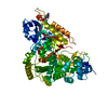
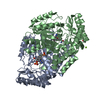
| Title | Crystal structure of 7,8-diaminopelargonic acid synthase (BioA) from Mycobacterium tuberculosis, complexed with a fragment hit | ||||||
 Components Components | Adenosylmethionine-8-amino-7-oxononanoate aminotransferase | ||||||
 Keywords Keywords | transferase/transferase inhibitor / Inhibitor Complex Transaminase PLP / transferase-transferase inhibitor complex | ||||||
| Function / homology |  Function and homology information Function and homology information adenosylmethionine-8-amino-7-oxononanoate transaminase / adenosylmethionine-8-amino-7-oxononanoate transaminase activity / dethiobiotin synthase activity / biotin biosynthetic process / pyridoxal phosphate binding / cytoplasm adenosylmethionine-8-amino-7-oxononanoate transaminase / adenosylmethionine-8-amino-7-oxononanoate transaminase activity / dethiobiotin synthase activity / biotin biosynthetic process / pyridoxal phosphate binding / cytoplasmSimilarity search - Function | ||||||
| Biological species |   Mycobacterium tuberculosis (bacteria) Mycobacterium tuberculosis (bacteria) | ||||||
| Method | X-RAY DIFFRACTION / MOLECULAR REPLACEMENT / Resolution: 2.5 Å | ||||||
 Authors Authors | Finzel, B.C. / Dai, D. / Geders, T.W. | ||||||
 Citation Citation | Journal: J.Med.Chem. / Year: 2015 Title: Fragment-Based Exploration of Binding Site Flexibility in Mycobacterium tuberculosis BioA. Authors: Dai, R. / Geders, T.W. / Liu, F. / Park, S.W. / Schnappinger, D. / Aldrich, C.C. / Finzel, B.C. | ||||||
| History |
|
- Structure visualization
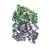
| Structure viewer | Molecule: MolmilJmol/JSmol |
|---|
- Downloads & links
-Download
| PDBx/mmCIF format | 4wya.cif.gz | 318.7 KB | Display | PDBx/mmCIF format |
|---|---|---|---|---|
| PDB format | pdb4wya.ent.gz | 257.3 KB | Display | PDB format |
| PDBx/mmJSON format | 4wya.json.gz | Tree view | PDBx/mmJSON format | |
| Others |  Other downloads Other downloads |
-Validation report
| Arichive directory | https://data.pdbj.org/pub/pdb/validation_reports/wy/4wyaftp://data.pdbj.org/pub/pdb/validation_reports/wy/4wya | HTTPS FTP |
|---|
-Related structure data
| Related structure data |  4wycC  4wydC  4wyeC  4wyfC  4wygC  4xewC  3tftS C: citing same article ( S: Starting model for refinement |
|---|---|
| Similar structure data |

-Links
 PDBj
PDBj- Assembly
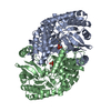
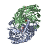
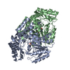
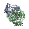
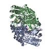
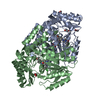
| Deposited unit | 
| ||||||||
|---|---|---|---|---|---|---|---|---|---|
| 1 | 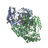
| ||||||||
| 2 | 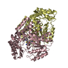
| ||||||||
| Unit cell |
|
-Components
| #1: Protein | Mass: 48536.520 Da / Num. of mol.: 4 Source method: isolated from a genetically manipulated source Source: (gene. exp.) Mycobacterium tuberculosis (strain ATCC 25618 / H37Rv) (bacteria)Strain: ATCC 25618 / H37Rv / Gene: bioA, Rv1568, MTCY336.35c / Production host: Escherichia coli (E. coli)References: UniProt: P9WQ81, adenosylmethionine-8-amino-7-oxononanoate transaminase#2: Chemical | ChemComp-PLP / Pyridoxal phosphate  Mass: 247.142 Da / Num. of mol.: 4 / Source method: obtained synthetically / Formula: C8H10NO6P Mass: 247.142 Da / Num. of mol.: 4 / Source method: obtained synthetically / Formula: C8H10NO6P#3: Chemical |   Mass: 204.248 Da / Num. of mol.: 2 / Source method: obtained synthetically / Formula: C10H8N2OS Mass: 204.248 Da / Num. of mol.: 2 / Source method: obtained synthetically / Formula: C10H8N2OS#4: Water | ChemComp-HOH / | Water Mass: 18.015 Da / Num. of mol.: 114 / Source method: isolated from a natural source / Formula: H2O Mass: 18.015 Da / Num. of mol.: 114 / Source method: isolated from a natural source / Formula: H2O |
|---|
-Experimental details
-Experiment
| Experiment | Method: X-RAY DIFFRACTION / Number of used crystals: 1 |
|---|
- Sample preparation
| Crystal | Density Matthews: 2.17 Å3/Da / Density % sol: 43.4 % |
|---|---|
| Crystal grow | Temperature: 293.15 K / Method: vapor diffusion, hanging drop / pH: 7.5 Details: Protein: 25 mM HEPES pH 7.5, 50 mM NaCl, 0.1 mM TCEP Reservoir:10% PEG 8000, 0.1M magnesium chloride, 0.1M HEPES pH 7.5 Cryo: 15% PEG 400 in reservoir solution, VAPOR DIFFUSION, HANGING DROP, temperature 293.15K |
-Data collection
| Diffraction | Mean temperature: 100 K |
|---|---|
| Diffraction source | Source: ROTATING ANODE / Type: RIGAKU FR-E+ DW / Wavelength: 1.54 Å |
| Detector | Type: RIGAKU SATURN 944+ / Detector: CCD / Date: Apr 4, 2013 |
| Radiation | Protocol: SINGLE WAVELENGTH / Monochromatic (M) / Laue (L): M / Scattering type: x-ray |
| Radiation wavelength | Wavelength: 1.54 Å / Relative weight: 1 |
| Reflection | Resolution: 2.5→29.866 Å / Num. obs: 50047 / % possible obs: 65 % / Redundancy: 1.92 % / Biso Wilson estimate: 38.2 Å2 / Rmerge(I) obs: 0.146 / Χ2: 0.95 / Net I/σ(I): 4.1 / Num. measured all: 136210 / Scaling rejects: 1022 |
| Reflection shell | Highest resolution: 2.5 Å |
- Processing
| Software |
| |||||||||||||||||||||||||||||||||||||||||||||||||||||||||||||||||||||||||||||||||||||||||||||||||||||||||||||||||||||||||||||||||||||
|---|---|---|---|---|---|---|---|---|---|---|---|---|---|---|---|---|---|---|---|---|---|---|---|---|---|---|---|---|---|---|---|---|---|---|---|---|---|---|---|---|---|---|---|---|---|---|---|---|---|---|---|---|---|---|---|---|---|---|---|---|---|---|---|---|---|---|---|---|---|---|---|---|---|---|---|---|---|---|---|---|---|---|---|---|---|---|---|---|---|---|---|---|---|---|---|---|---|---|---|---|---|---|---|---|---|---|---|---|---|---|---|---|---|---|---|---|---|---|---|---|---|---|---|---|---|---|---|---|---|---|---|---|---|---|
| Refinement | Method to determine structure: MOLECULAR REPLACEMENT Starting model: 3TFT Resolution: 2.5→29.866 Å / FOM work R set: 0.6997 / SU ML: 0.43 / Cross valid method: FREE R-VALUE / σ(F): 1.33 / Phase error: 35.91 / Stereochemistry target values: ML
| |||||||||||||||||||||||||||||||||||||||||||||||||||||||||||||||||||||||||||||||||||||||||||||||||||||||||||||||||||||||||||||||||||||
| Solvent computation | Shrinkage radii: 0.9 Å / VDW probe radii: 1.11 Å / Solvent model: FLAT BULK SOLVENT MODEL | |||||||||||||||||||||||||||||||||||||||||||||||||||||||||||||||||||||||||||||||||||||||||||||||||||||||||||||||||||||||||||||||||||||
| Displacement parameters | Biso max: 92.37 Å2 / Biso mean: 43.1 Å2 / Biso min: 21.05 Å2 | |||||||||||||||||||||||||||||||||||||||||||||||||||||||||||||||||||||||||||||||||||||||||||||||||||||||||||||||||||||||||||||||||||||
| Refinement step | Cycle: final / Resolution: 2.5→29.866 Å
| |||||||||||||||||||||||||||||||||||||||||||||||||||||||||||||||||||||||||||||||||||||||||||||||||||||||||||||||||||||||||||||||||||||
| Refine LS restraints |
| |||||||||||||||||||||||||||||||||||||||||||||||||||||||||||||||||||||||||||||||||||||||||||||||||||||||||||||||||||||||||||||||||||||
| LS refinement shell | Refine-ID: X-RAY DIFFRACTION / Total num. of bins used: 18
|
