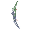
 Movie
Movie Controller


+ Open data

- Basic information
| Entry | Database: PDB / ID: 4wpe | ||||||
|---|---|---|---|---|---|---|---|
| Title | Crystal Structure of Hof1p F-BAR domain | ||||||
 Components Components | Cytokinesis protein 2 | ||||||
 Keywords Keywords | PROTEIN BINDING / F-BAR domain / membrane | ||||||
| Function / homology |  Function and homology information Function and homology informationprimary cell septum biogenesis / protein localization to cell division site / negative regulation of formin-nucleated actin cable assembly / HICS complex / regulation of mitotic actomyosin contractile ring contraction / : / mitotic actomyosin contractile ring maturation / MIH complex / protein localization to cell division site involved in mitotic actomyosin contractile ring assembly / Clathrin-mediated endocytosis ...primary cell septum biogenesis / protein localization to cell division site / negative regulation of formin-nucleated actin cable assembly / HICS complex / regulation of mitotic actomyosin contractile ring contraction / : / mitotic actomyosin contractile ring maturation / MIH complex / protein localization to cell division site involved in mitotic actomyosin contractile ring assembly / Clathrin-mediated endocytosis / mitotic actomyosin contractile ring, proximal layer / myosin II heavy chain binding / cellular bud neck septin ring / mitotic actomyosin contractile ring assembly / cellular bud neck contractile ring / site of polarized growth / actomyosin contractile ring / cellular bud neck / cell division site / mitotic cytokinesis / cytoskeletal protein binding / phospholipid binding / cell cortex / cytoskeleton / protein-containing complex binding / identical protein binding / plasma membrane / cytoplasmSimilarity search - Function | ||||||
| Biological species |  Saccharomyces cerevisiae (brewer's yeast) Saccharomyces cerevisiae (brewer's yeast) | ||||||
| Method | X-RAY DIFFRACTION / SYNCHROTRON / Resolution: 2.7 Å | ||||||
 Authors Authors | Lemmon, M.A. / Moravcevic, K. | ||||||
 Citation Citation | Journal: Structure / Year: 2015 Title: Comparison of Saccharomyces cerevisiae F-BAR Domain Structures Reveals a Conserved Inositol Phosphate Binding Site. Authors: Moravcevic, K. / Alvarado, D. / Schmitz, K.R. / Kenniston, J.A. / Mendrola, J.M. / Ferguson, K.M. / Lemmon, M.A. | ||||||
| History |
|
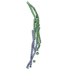
- Structure visualization
| Structure viewer | Molecule: MolmilJmol/JSmol |
|---|
- Downloads & links
-Download
| PDBx/mmCIF format | 4wpe.cif.gz | 125 KB | Display | PDBx/mmCIF format |
|---|---|---|---|---|
| PDB format | pdb4wpe.ent.gz | 97.1 KB | Display | PDB format |
| PDBx/mmJSON format | 4wpe.json.gz | Tree view | PDBx/mmJSON format | |
| Others |  Other downloads Other downloads |
-Validation report
| Arichive directory | https://data.pdbj.org/pub/pdb/validation_reports/wp/4wpeftp://data.pdbj.org/pub/pdb/validation_reports/wp/4wpe | HTTPS FTP |
|---|
-Related structure data


-Links
 PDBj
PDBj

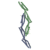
- Assembly
| Deposited unit | 
| ||||||||
|---|---|---|---|---|---|---|---|---|---|
| 1 | 
| ||||||||
| Unit cell |
| ||||||||
| Components on special symmetry positions |
|
-Components
| #1: Protein | / Homolog of CDC15 protein 1 Mass: 36095.098 Da / Num. of mol.: 1 Source method: isolated from a genetically manipulated source Source: (gene. exp.) Saccharomyces cerevisiae (brewer's yeast)Strain: ATCC 204508 / S288c / Gene: HOF1, CYK2, YMR032W, YM9973.05 / Production host:  Escherichia coli (E. coli) / Strain (production host): B834 / References: UniProt: Q05080 Escherichia coli (E. coli) / Strain (production host): B834 / References: UniProt: Q05080 |
|---|---|
| #2: Water | ChemComp-HOH / Water Mass: 18.015 Da / Num. of mol.: 40 / Source method: isolated from a natural source / Formula: H2O Mass: 18.015 Da / Num. of mol.: 40 / Source method: isolated from a natural source / Formula: H2O |
-Experimental details
-Experiment
| Experiment | Method: X-RAY DIFFRACTION |
|---|
- Sample preparation
| Crystal | Density Matthews: 2.94 Å3/Da / Density % sol: 58.17 % |
|---|---|
| Crystal grow | Temperature: 294 K / Method: vapor diffusion, hanging drop / pH: 5.5 Details: 0.1 M Na citrate, pH 5.5, containing 0.1 M ammonium acetate, and 5-7% (w/v) PEG3350 |
-Data collection
| Diffraction | Mean temperature: 100 K |
|---|---|
| Diffraction source | Source: SYNCHROTRON / Site: APS  / Beamline: 23-ID-B / Wavelength: 0.97949 Å / Beamline: 23-ID-B / Wavelength: 0.97949 Å |
| Detector | Type: MARMOSAIC 300 mm CCD / Detector: CCD / Date: Aug 5, 2007 |
| Radiation | Protocol: MAD / Monochromatic (M) / Laue (L): M / Scattering type: x-ray |
| Radiation wavelength | Wavelength: 0.97949 Å / Relative weight: 1 |
| Reflection | Resolution: 2.7→50 Å / Num. obs: 11489 / % possible obs: 98.9 % / Redundancy: 7.2 % / Net I/σ(I): 33.9 |
- Processing
| Software | Name: PHENIX / Version: (phenix.refine: 1.9_1692) / Classification: refinement | ||||||||||||||||||||||||||||||||||||||||
|---|---|---|---|---|---|---|---|---|---|---|---|---|---|---|---|---|---|---|---|---|---|---|---|---|---|---|---|---|---|---|---|---|---|---|---|---|---|---|---|---|---|
| Refinement | Resolution: 2.7→39.908 Å / SU ML: 0.18 / Cross valid method: FREE R-VALUE / σ(F): 1.35 / Phase error: 31.32 / Stereochemistry target values: ML
| ||||||||||||||||||||||||||||||||||||||||
| Solvent computation | Shrinkage radii: 0.9 Å / VDW probe radii: 1.11 Å / Solvent model: FLAT BULK SOLVENT MODEL | ||||||||||||||||||||||||||||||||||||||||
| Refinement step | Cycle: LAST / Resolution: 2.7→39.908 Å
| ||||||||||||||||||||||||||||||||||||||||
| Refine LS restraints |
| ||||||||||||||||||||||||||||||||||||||||
| LS refinement shell |
| ||||||||||||||||||||||||||||||||||||||||
| Refinement TLS params. | Method: refined / Origin x: 23.3817 Å / Origin y: 39.7422 Å / Origin z: 61.4186 Å
| ||||||||||||||||||||||||||||||||||||||||
| Refinement TLS group | Selection details: all |
