 Movie
Movie Controller

[English] 日本語
 Yorodumi
- PDB-4wpc: Crystal structure of Rgd1p F-BAR domain in complex with inositol ... -
+ Open data

- Basic information
| Entry | Database: PDB / ID: 4wpc | ||||||
|---|---|---|---|---|---|---|---|
| Title | Crystal structure of Rgd1p F-BAR domain in complex with inositol phosphate | ||||||
 Components Components | RHO GTPase-activating protein RGD1 | ||||||
 Keywords Keywords | LIPID BINDING PROTEIN /  F-BAR domain / phospholipid binding / PROTEIN BINDING F-BAR domain / phospholipid binding / PROTEIN BINDING | ||||||
| Function / homology |  Function and homology information: / : / RHOF GTPase cycle / CDC42 GTPase cycle / RHOD GTPase cycle / RHOV GTPase cycle / RHOA GTPase cycle / cellular bud / prospore membrane / actin cortical patch ...: / : / RHOF GTPase cycle / CDC42 GTPase cycle / RHOD GTPase cycle / RHOV GTPase cycle / RHOA GTPase cycle / cellular bud / prospore membrane / actin cortical patch / phosphatidylinositol-5-phosphate binding / mating projection tip / phosphatidylinositol-3-phosphate binding / response to acidic pH / phosphatidylinositol-4-phosphate binding / phosphatidylinositol-3,5-bisphosphate binding / response to osmotic stress / Neutrophil degranulation / phosphatidylinositol-4,5-bisphosphate binding / GTPase activator activity / signal transduction / identical protein binding / cytoplasm Function and homology information: / : / RHOF GTPase cycle / CDC42 GTPase cycle / RHOD GTPase cycle / RHOV GTPase cycle / RHOA GTPase cycle / cellular bud / prospore membrane / actin cortical patch ...: / : / RHOF GTPase cycle / CDC42 GTPase cycle / RHOD GTPase cycle / RHOV GTPase cycle / RHOA GTPase cycle / cellular bud / prospore membrane / actin cortical patch / phosphatidylinositol-5-phosphate binding / mating projection tip / phosphatidylinositol-3-phosphate binding / response to acidic pH / phosphatidylinositol-4-phosphate binding / phosphatidylinositol-3,5-bisphosphate binding / response to osmotic stress / Neutrophil degranulation / phosphatidylinositol-4,5-bisphosphate binding / GTPase activator activity / signal transduction / identical protein binding / cytoplasmSimilarity search - Function | ||||||
| Biological species |  Saccharomyces cerevisiae (brewer's yeast) Saccharomyces cerevisiae (brewer's yeast) | ||||||
| Method | X-RAY DIFFRACTION / SYNCHROTRON / MOLECULAR REPLACEMENT / Resolution: 3.34 Å | ||||||
 Authors Authors | Moravcevic, K. / Lemmon, M.A. | ||||||
 Citation Citation | Journal: Structure / Year: 2015 Title: Comparison of Saccharomyces cerevisiae F-BAR Domain Structures Reveals a Conserved Inositol Phosphate Binding Site. Authors: Moravcevic, K. / Alvarado, D. / Schmitz, K.R. / Kenniston, J.A. / Mendrola, J.M. / Ferguson, K.M. / Lemmon, M.A. | ||||||
| History |
|
- Structure visualization
| Structure viewer | Molecule: MolmilJmol/JSmol |
|---|
- Downloads & links
-Download
| PDBx/mmCIF format | 4wpc.cif.gz | 210.8 KB | Display | PDBx/mmCIF format |
|---|---|---|---|---|
| PDB format | pdb4wpc.ent.gz | 170.2 KB | Display | PDB format |
| PDBx/mmJSON format | 4wpc.json.gz | Tree view | PDBx/mmJSON format | |
| Others |  Other downloads Other downloads |
-Validation report
| Arichive directory | https://data.pdbj.org/pub/pdb/validation_reports/wp/4wpcftp://data.pdbj.org/pub/pdb/validation_reports/wp/4wpc | HTTPS FTP |
|---|
-Related structure data
| Related structure data |  4wpeSC S: Starting model for refinement C: citing same article ( |
|---|---|
| Similar structure data |


-Links
 PDBj
PDBj


- Assembly
| Deposited unit | 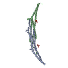
| ||||||||
|---|---|---|---|---|---|---|---|---|---|
| 1 |
| ||||||||
| Unit cell |
|
-Components
| #1: Protein | Mass: 36879.336 Da / Num. of mol.: 2 Source method: isolated from a genetically manipulated source Source: (gene. exp.) Saccharomyces cerevisiae (brewer's yeast)Strain: ATCC 204508 / S288c / Gene: RGD1, YBR260C, YBR1728 / Production host:  Escherichia coli (E. coli) / Strain (production host): BL21DE3 / References: UniProt: P38339 Escherichia coli (E. coli) / Strain (production host): BL21DE3 / References: UniProt: P38339#2: Chemical | Phytic acid  Mass: 660.035 Da / Num. of mol.: 2 / Source method: obtained synthetically / Formula: C6H18O24P6 Mass: 660.035 Da / Num. of mol.: 2 / Source method: obtained synthetically / Formula: C6H18O24P6#3: Water | ChemComp-HOH / | Water Mass: 18.015 Da / Num. of mol.: 8 / Source method: isolated from a natural source / Formula: H2O Mass: 18.015 Da / Num. of mol.: 8 / Source method: isolated from a natural source / Formula: H2O |
|---|
-Experimental details
-Experiment
| Experiment | Method: X-RAY DIFFRACTION |
|---|
- Sample preparation
| Crystal | Density Matthews: 4.61 Å3/Da / Density % sol: 73.3 % |
|---|---|
| Crystal grow | Temperature: 294 K / Method: vapor diffusion, hanging drop Details: 0.1 M citrate, pH 5.5, containing 0.1-0.2 M (NH4)2SO4 plus 10-20% (w/v) PEG3350 PH range: 5.5 |
-Data collection
| Diffraction | Mean temperature: 100 K |
|---|---|
| Diffraction source | Source: SYNCHROTRON / Site: CHESS  / Beamline: F1 / Wavelength: 0.9179 Å / Beamline: F1 / Wavelength: 0.9179 Å |
| Detector | Type: ADSC QUANTUM 270 / Detector: CCD / Date: Nov 9, 2007 |
| Radiation | Protocol: SINGLE WAVELENGTH / Monochromatic (M) / Laue (L): M / Scattering type: x-ray |
| Radiation wavelength | Wavelength: 0.9179 Å / Relative weight: 1 |
| Reflection | Resolution: 3.3→50 Å / Num. obs: 19341 / % possible obs: 99.7 % / Redundancy: 4.2 % / Net I/σ(I): 14 |
- Processing
| Software |
| ||||||||||||||||||||||||||||||||||||||||||||||||||||||||
|---|---|---|---|---|---|---|---|---|---|---|---|---|---|---|---|---|---|---|---|---|---|---|---|---|---|---|---|---|---|---|---|---|---|---|---|---|---|---|---|---|---|---|---|---|---|---|---|---|---|---|---|---|---|---|---|---|---|
| Refinement | Method to determine structure: MOLECULAR REPLACEMENT Starting model: PDB ENTRY 4WPE Resolution: 3.34→30.773 Å / SU ML: 0.47 / Cross valid method: FREE R-VALUE / σ(F): 1.33 / Phase error: 30.01 / Stereochemistry target values: ML
| ||||||||||||||||||||||||||||||||||||||||||||||||||||||||
| Solvent computation | Shrinkage radii: 0.9 Å / VDW probe radii: 1.11 Å / Solvent model: FLAT BULK SOLVENT MODEL | ||||||||||||||||||||||||||||||||||||||||||||||||||||||||
| Refinement step | Cycle: LAST / Resolution: 3.34→30.773 Å
| ||||||||||||||||||||||||||||||||||||||||||||||||||||||||
| Refine LS restraints |
| ||||||||||||||||||||||||||||||||||||||||||||||||||||||||
| LS refinement shell |
|
