 Movie
Movie Controller

[English] 日本語
 Yorodumi
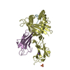
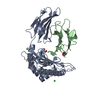
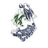
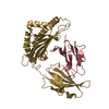
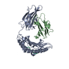
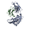
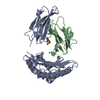
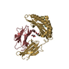
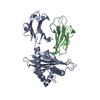
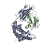
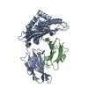
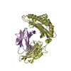
- PDB-4wdi: Weak TCR binding to an unstable insulin epitope drives type 1 diabetes -
+ Open data

- Basic information
| Entry | Database: PDB / ID: 4wdi | ||||||
|---|---|---|---|---|---|---|---|
| Title | Weak TCR binding to an unstable insulin epitope drives type 1 diabetes | ||||||
 Components Components |
| ||||||
 Keywords Keywords |  IMMUNE SYSTEM / Immunoglobulin / H-2Kd / Type 1 Diabetes IMMUNE SYSTEM / Immunoglobulin / H-2Kd / Type 1 Diabetes | ||||||
| Function / homology |  Function and homology information Function and homology informationnegative regulation of NAD(P)H oxidase activity / negative regulation of glycogen catabolic process / regulation of cellular amino acid metabolic process / negative regulation of fatty acid metabolic process / negative regulation of feeding behavior / Signaling by Insulin receptor / nitric oxide-cGMP-mediated signaling / IRS activation / Insulin processing / regulation of protein secretion ...negative regulation of NAD(P)H oxidase activity / negative regulation of glycogen catabolic process / regulation of cellular amino acid metabolic process / negative regulation of fatty acid metabolic process / negative regulation of feeding behavior / Signaling by Insulin receptor / nitric oxide-cGMP-mediated signaling / IRS activation / Insulin processing / regulation of protein secretion / positive regulation of peptide hormone secretion / positive regulation of respiratory burst / antigen processing and presentation of exogenous peptide antigen via MHC class I / Regulation of gene expression in beta cells / negative regulation of acute inflammatory response / inner ear development / alpha-beta T cell activation / antigen processing and presentation of endogenous peptide antigen via MHC class I via ER pathway, TAP-dependent / negative regulation of respiratory burst involved in inflammatory response / positive regulation of dendritic spine maintenance / positive regulation of glycogen biosynthetic process / Synthesis, secretion, and deacylation of Ghrelin / negative regulation of protein secretion / positive regulation of nitric oxide mediated signal transduction / fatty acid homeostasis / regulation of protein localization to plasma membrane / Signal attenuation / FOXO-mediated transcription of oxidative stress, metabolic and neuronal genes / negative regulation of lipid catabolic process / beta-2-microglobulin binding / negative regulation of gluconeogenesis / COPI-mediated anterograde transport / positive regulation of lipid biosynthetic process / negative regulation of oxidative stress-induced intrinsic apoptotic signaling pathway / negative regulation of reactive oxygen species biosynthetic process / positive regulation of insulin receptor signaling pathway / transport vesicle / positive regulation of protein autophosphorylation / Insulin receptor recycling / insulin-like growth factor receptor binding / neuron projection maintenance / NPAS4 regulates expression of target genes / positive regulation of protein metabolic process / positive regulation of brown fat cell differentiation / endoplasmic reticulum-Golgi intermediate compartment membrane / positive regulation of glycolytic process / activation of protein kinase B activity / positive regulation of mitotic nuclear division / Insulin receptor signalling cascade / antigen processing and presentation of endogenous peptide antigen via MHC class I via ER pathway, TAP-independent / antigen processing and presentation of endogenous peptide antigen via MHC class Ib / positive regulation of ferrous iron binding / positive regulation of transferrin receptor binding / positive regulation of receptor binding / early endosome lumen / Nef mediated downregulation of MHC class I complex cell surface expression / DAP12 interactions / negative regulation of receptor binding / positive regulation of cytokine production / positive regulation of long-term synaptic potentiation / Regulation of insulin secretion / acute-phase response / endosome lumen / positive regulation of protein secretion / positive regulation of glucose import / positive regulation of nitric-oxide synthase activity / lumenal side of endoplasmic reticulum membrane / positive regulation of cell differentiation / negative regulation of proteolysis / Endosomal/Vacuolar pathway / regulation of transmembrane transporter activity / Antigen Presentation: Folding, assembly and peptide loading of class I MHC / peptide binding / wound healing / cellular response to iron(III) ion / antigen processing and presentation of exogenous protein antigen via MHC class Ib, TAP-dependent / regulation of synaptic plasticity / negative regulation of forebrain neuron differentiation / insulin receptor binding / ER to Golgi transport vesicle membrane / regulation of erythrocyte differentiation / peptide antigen assembly with MHC class I protein complex / response to molecule of bacterial origin / regulation of iron ion transport / MHC class I peptide loading complex / HFE-transferrin receptor complex / negative regulation of protein catabolic process / T cell mediated cytotoxicity / hormone activity / cellular response to iron ion / antigen processing and presentation of endogenous peptide antigen via MHC class I / positive regulation of T cell cytokine production / cognition / MHC class I protein complex / multicellular organismal-level iron ion homeostasis / positive regulation of neuron projection development / negative regulation of neurogenesis / peptide antigen assembly with MHC class II protein complex / positive regulation of receptor-mediated endocytosis / Golgi lumenSimilarity search - Function | ||||||
| Biological species |  Mus musculus (house mouse) Mus musculus (house mouse) Homo sapiens (human) Homo sapiens (human) | ||||||
| Method | X-RAY DIFFRACTION / SYNCHROTRON / MOLECULAR REPLACEMENT / molecular replacement / Resolution: 2.313 Å | ||||||
| Model details | G9G in H-2Kd, Triclinic form | ||||||
 Authors Authors | Rizkallah, P.J. / Cole, D.K. | ||||||
 Citation Citation | Journal: J.Biol.Chem. / Year: 2015 Title: Distortion of the Major Histocompatibility Complex Class I Binding Groove to Accommodate an Insulin-derived 10-Mer Peptide. Authors: Motozono, C. / Pearson, J.A. / De Leenheer, E. / Rizkallah, P.J. / Beck, K. / Trimby, A. / Sewell, A.K. / Wong, F.S. / Cole, D.K. | ||||||
| History |
|
- Structure visualization
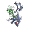
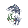
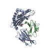
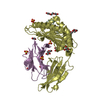
| Structure viewer | Molecule: MolmilJmol/JSmol |
|---|
- Downloads & links
-Download
| PDBx/mmCIF format | 4wdi.cif.gz | 329 KB | Display | PDBx/mmCIF format |
|---|---|---|---|---|
| PDB format | pdb4wdi.ent.gz | 280.2 KB | Display | PDB format |
| PDBx/mmJSON format | 4wdi.json.gz | Tree view | PDBx/mmJSON format | |
| Others |  Other downloads Other downloads |
-Validation report
| Arichive directory | https://data.pdbj.org/pub/pdb/validation_reports/wd/4wdiftp://data.pdbj.org/pub/pdb/validation_reports/wd/4wdi | HTTPS FTP |
|---|
-Related structure data
| Related structure data | 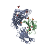 4z76C 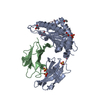 4z78C  4wdj 4wdk 4wdl C: citing same article ( |
|---|---|
| Similar structure data |


-Links
 PDBj
PDBj


- Assembly
| Deposited unit | 
| ||||||||
|---|---|---|---|---|---|---|---|---|---|
| 1 | 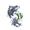
| ||||||||
| 2 | 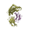
| ||||||||
| Unit cell |
| ||||||||
| Details | Chains A, B and C form one biological entity. / Chains D,E,F form one biological entity. |
-Components
-Protein , 2 types, 4 molecules ADBE
| #1: Protein | Mass: 32353.016 Da / Num. of mol.: 2 / Fragment: H-2Kd MHC, residues 22-296 Source method: isolated from a genetically manipulated source Source: (gene. exp.) Mus musculus (house mouse) / Gene: H2-K1, H2-K / Plasmid: pGMT7 / Production host:  Escherichia coli (E. coli) / Variant (production host): rosetta / References: UniProt: P01902*PLUS Escherichia coli (E. coli) / Variant (production host): rosetta / References: UniProt: P01902*PLUS#2: Protein | Beta-2 microglobulinMass: 11879.356 Da / Num. of mol.: 2 / Fragment: Human beta2-microglobulin, residues 21-219 Source method: isolated from a genetically manipulated source Source: (gene. exp.) Homo sapiens (human) / Gene: B2M, CDABP0092, HDCMA22P / Plasmid: pGMT7 / Production host: Escherichia coli BL21(DE3) (bacteria) / Variant (production host): rosetta / References: UniProt: P61769 |
|---|
-Protein/peptide , 1 types, 2 molecules CF
| #3: Protein/peptide | Mass: 1010.190 Da / Num. of mol.: 2 Source method: isolated from a genetically manipulated source Source: (gene. exp.) Homo sapiens (human) / Gene: INS / Production host: synthetic construct (others) / References: UniProt: P01308 |
|---|
-Non-polymers , 3 types, 165 molecules 


| #4: Chemical | ChemComp-EDO / Ethylene glycol Mass: 62.068 Da / Num. of mol.: 1 / Source method: obtained synthetically / Formula: C2H6O2 Mass: 62.068 Da / Num. of mol.: 1 / Source method: obtained synthetically / Formula: C2H6O2 | ||
|---|---|---|---|
| #5: Chemical | ChemComp-SO4 / Sulfate Mass: 96.063 Da / Num. of mol.: 4 / Source method: obtained synthetically / Formula: SO4 Mass: 96.063 Da / Num. of mol.: 4 / Source method: obtained synthetically / Formula: SO4#6: Water | ChemComp-HOH / | WaterMass: 18.015 Da / Num. of mol.: 160 / Source method: isolated from a natural source / Formula: H2O |
-Experimental details
-Experiment
| Experiment | Method: X-RAY DIFFRACTION / Number of used crystals: 1 |
|---|
- Sample preparation
| Crystal | Density Matthews: 2.18 Å3/Da / Density % sol: 43.64 % |
|---|---|
| Crystal grow | Temperature: 291 K / Method: vapor diffusion, sitting drop / pH: 8 Details: 20% PEG 3350, 0.2 M Sodium malonate, and 0.1 M Bis-Tris Propane, pH 6.5 |
-Data collection
| Diffraction | Mean temperature: 100 K | ||||||||||||||||||||||||||||||||||||||||||||||||||||||||||||||||||||||||||||||||||||||||||||||||||||||||||||||||||||||||||||||||||||||||||||||||||||||||||||||||||||||||||||||||||||||||||||||||||||||||||||||||||
|---|---|---|---|---|---|---|---|---|---|---|---|---|---|---|---|---|---|---|---|---|---|---|---|---|---|---|---|---|---|---|---|---|---|---|---|---|---|---|---|---|---|---|---|---|---|---|---|---|---|---|---|---|---|---|---|---|---|---|---|---|---|---|---|---|---|---|---|---|---|---|---|---|---|---|---|---|---|---|---|---|---|---|---|---|---|---|---|---|---|---|---|---|---|---|---|---|---|---|---|---|---|---|---|---|---|---|---|---|---|---|---|---|---|---|---|---|---|---|---|---|---|---|---|---|---|---|---|---|---|---|---|---|---|---|---|---|---|---|---|---|---|---|---|---|---|---|---|---|---|---|---|---|---|---|---|---|---|---|---|---|---|---|---|---|---|---|---|---|---|---|---|---|---|---|---|---|---|---|---|---|---|---|---|---|---|---|---|---|---|---|---|---|---|---|---|---|---|---|---|---|---|---|---|---|---|---|---|---|---|---|---|
| Diffraction source | Source: SYNCHROTRON / Site: Diamond  / Beamline: I04-1 / Wavelength: 0.917 Å / Beamline: I04-1 / Wavelength: 0.917 Å | ||||||||||||||||||||||||||||||||||||||||||||||||||||||||||||||||||||||||||||||||||||||||||||||||||||||||||||||||||||||||||||||||||||||||||||||||||||||||||||||||||||||||||||||||||||||||||||||||||||||||||||||||||
| Detector | Type: DECTRIS PILATUS 2M / Detector: PIXEL / Date: Feb 9, 2012 / Details: mirrors | ||||||||||||||||||||||||||||||||||||||||||||||||||||||||||||||||||||||||||||||||||||||||||||||||||||||||||||||||||||||||||||||||||||||||||||||||||||||||||||||||||||||||||||||||||||||||||||||||||||||||||||||||||
| Radiation | Protocol: SINGLE WAVELENGTH / Monochromatic (M) / Laue (L): M / Scattering type: x-ray | ||||||||||||||||||||||||||||||||||||||||||||||||||||||||||||||||||||||||||||||||||||||||||||||||||||||||||||||||||||||||||||||||||||||||||||||||||||||||||||||||||||||||||||||||||||||||||||||||||||||||||||||||||
| Radiation wavelength |
| ||||||||||||||||||||||||||||||||||||||||||||||||||||||||||||||||||||||||||||||||||||||||||||||||||||||||||||||||||||||||||||||||||||||||||||||||||||||||||||||||||||||||||||||||||||||||||||||||||||||||||||||||||
| Reflection | Resolution: 2.313→29.518 Å / Num. all: 30132 / Num. obs: 30132 / % possible obs: 90.2 % / Redundancy: 2.1 % / Rpim(I) all: 0.081 / Rrim(I) all: 0.12 / Rsym value: 0.068 / Net I/av σ(I): 10 / Net I/σ(I): 8.6 / Num. measured all: 64672 | ||||||||||||||||||||||||||||||||||||||||||||||||||||||||||||||||||||||||||||||||||||||||||||||||||||||||||||||||||||||||||||||||||||||||||||||||||||||||||||||||||||||||||||||||||||||||||||||||||||||||||||||||||
| Reflection shell | Diffraction-ID: 1 / Rejects: 0
|
-Phasing
| Phasing | Method: molecular replacement |
|---|
- Processing
| Software |
| |||||||||||||||||||||||||||||||||||||||||||||||||||||||||||||||||||||||||||||||||||||||||||||||||||||||||||||||||||||||||||||||||||||||||||||||||||||||||||||||||||||||||||||||
|---|---|---|---|---|---|---|---|---|---|---|---|---|---|---|---|---|---|---|---|---|---|---|---|---|---|---|---|---|---|---|---|---|---|---|---|---|---|---|---|---|---|---|---|---|---|---|---|---|---|---|---|---|---|---|---|---|---|---|---|---|---|---|---|---|---|---|---|---|---|---|---|---|---|---|---|---|---|---|---|---|---|---|---|---|---|---|---|---|---|---|---|---|---|---|---|---|---|---|---|---|---|---|---|---|---|---|---|---|---|---|---|---|---|---|---|---|---|---|---|---|---|---|---|---|---|---|---|---|---|---|---|---|---|---|---|---|---|---|---|---|---|---|---|---|---|---|---|---|---|---|---|---|---|---|---|---|---|---|---|---|---|---|---|---|---|---|---|---|---|---|---|---|---|---|---|---|
| Refinement | Method to determine structure: MOLECULAR REPLACEMENT / Resolution: 2.313→29.518 Å / Cor.coef. Fo:Fc: 0.941 / Cor.coef. Fo:Fc free: 0.895 / WRfactor Rfree: 0.2791 / WRfactor Rwork: 0.2004 / FOM work R set: 0.8015 / SU B: 19.774 / SU ML: 0.235 / SU R Cruickshank DPI: 0.861 / SU Rfree: 0.3211 / Cross valid method: THROUGHOUT / σ(F): 0 / ESU R: 0.861 / ESU R Free: 0.321 / Stereochemistry target values: MAXIMUM LIKELIHOOD Details: HYDROGENS HAVE BEEN ADDED IN THE RIDING POSITIONS U VALUES : WITH TLS ADDED
| |||||||||||||||||||||||||||||||||||||||||||||||||||||||||||||||||||||||||||||||||||||||||||||||||||||||||||||||||||||||||||||||||||||||||||||||||||||||||||||||||||||||||||||||
| Solvent computation | Ion probe radii: 0.8 Å / Shrinkage radii: 0.8 Å / VDW probe radii: 1.4 Å / Solvent model: MASK | |||||||||||||||||||||||||||||||||||||||||||||||||||||||||||||||||||||||||||||||||||||||||||||||||||||||||||||||||||||||||||||||||||||||||||||||||||||||||||||||||||||||||||||||
| Displacement parameters | Biso max: 100.76 Å2 / Biso mean: 32.807 Å2 / Biso min: 11.48 Å2
| |||||||||||||||||||||||||||||||||||||||||||||||||||||||||||||||||||||||||||||||||||||||||||||||||||||||||||||||||||||||||||||||||||||||||||||||||||||||||||||||||||||||||||||||
| Refinement step | Cycle: final / Resolution: 2.313→29.518 Å
| |||||||||||||||||||||||||||||||||||||||||||||||||||||||||||||||||||||||||||||||||||||||||||||||||||||||||||||||||||||||||||||||||||||||||||||||||||||||||||||||||||||||||||||||
| Refine LS restraints |
| |||||||||||||||||||||||||||||||||||||||||||||||||||||||||||||||||||||||||||||||||||||||||||||||||||||||||||||||||||||||||||||||||||||||||||||||||||||||||||||||||||||||||||||||
| LS refinement shell | Resolution: 2.313→2.373 Å / Total num. of bins used: 20
| |||||||||||||||||||||||||||||||||||||||||||||||||||||||||||||||||||||||||||||||||||||||||||||||||||||||||||||||||||||||||||||||||||||||||||||||||||||||||||||||||||||||||||||||
| Refinement TLS params. | Method: refined / Refine-ID: X-RAY DIFFRACTION
| |||||||||||||||||||||||||||||||||||||||||||||||||||||||||||||||||||||||||||||||||||||||||||||||||||||||||||||||||||||||||||||||||||||||||||||||||||||||||||||||||||||||||||||||
| Refinement TLS group |
|
