 Movie
Movie Controller

[English] 日本語
 Yorodumi
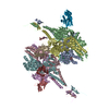
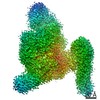
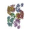
- PDB-4w8f: Crystal structure of the dynein motor domain in the AMPPNP-bound state -
+ Open data

- Basic information
| Entry | Database: PDB / ID: 4w8f | ||||||
|---|---|---|---|---|---|---|---|
| Title | Crystal structure of the dynein motor domain in the AMPPNP-bound state | ||||||
 Components Components | Dynein heavy chain lysozyme chimera | ||||||
 Keywords Keywords |  MOTOR PROTEIN / cytoplasmic dynein / microtubule / ATPase / AAA+ / AMPPNP MOTOR PROTEIN / cytoplasmic dynein / microtubule / ATPase / AAA+ / AMPPNP | ||||||
| Function / homology |  Function and homology informationkaryogamy / establishment of mitotic spindle localization / astral microtubule / nuclear migration along microtubule / minus-end-directed microtubule motor activity / cytoplasmic dynein complex / dynein light intermediate chain binding / spindle pole body / nuclear migration / dynein intermediate chain binding ...karyogamy / establishment of mitotic spindle localization / astral microtubule / nuclear migration along microtubule / minus-end-directed microtubule motor activity / cytoplasmic dynein complex / dynein light intermediate chain binding / spindle pole body / nuclear migration / dynein intermediate chain binding / mitotic sister chromatid segregation / establishment of mitotic spindle orientation / cytoplasmic microtubule / cytoplasmic microtubule organization / viral release from host cell by cytolysis / Neutrophil degranulation / peptidoglycan catabolic process / mitotic spindle organization / cell wall macromolecule catabolic process / lysozyme / lysozyme activity / cell cortex / host cell cytoplasm / defense response to bacterium / ATP hydrolysis activity / ATP binding / cytoplasm Function and homology informationkaryogamy / establishment of mitotic spindle localization / astral microtubule / nuclear migration along microtubule / minus-end-directed microtubule motor activity / cytoplasmic dynein complex / dynein light intermediate chain binding / spindle pole body / nuclear migration / dynein intermediate chain binding ...karyogamy / establishment of mitotic spindle localization / astral microtubule / nuclear migration along microtubule / minus-end-directed microtubule motor activity / cytoplasmic dynein complex / dynein light intermediate chain binding / spindle pole body / nuclear migration / dynein intermediate chain binding / mitotic sister chromatid segregation / establishment of mitotic spindle orientation / cytoplasmic microtubule / cytoplasmic microtubule organization / viral release from host cell by cytolysis / Neutrophil degranulation / peptidoglycan catabolic process / mitotic spindle organization / cell wall macromolecule catabolic process / lysozyme / lysozyme activity / cell cortex / host cell cytoplasm / defense response to bacterium / ATP hydrolysis activity / ATP binding / cytoplasmSimilarity search - Function | ||||||
| Biological species |  Saccharomyces cerevisiae (brewer's yeast) Saccharomyces cerevisiae (brewer's yeast) Enterobacteria phage T4 (virus) Enterobacteria phage T4 (virus) | ||||||
| Method | X-RAY DIFFRACTION / SYNCHROTRON / Resolution: 3.541 Å | ||||||
 Authors Authors | Cheng, H.-C. / Bhabha, G. / Zhang, N. / Vale, R.D. | ||||||
| Funding support |  United States, 1items United States, 1items
| ||||||
 Citation Citation | Journal: Cell / Year: 2014 Title: Allosteric communication in the dynein motor domain. Authors: Gira Bhabha / Hui-Chun Cheng / Nan Zhang / Arne Moeller / Maofu Liao / Jeffrey A Speir / Yifan Cheng / Ronald D Vale / Abstract: Dyneins power microtubule motility using ring-shaped, AAA-containing motor domains. Here, we report X-ray and electron microscopy (EM) structures of yeast dynein bound to different ATP analogs, which ...Dyneins power microtubule motility using ring-shaped, AAA-containing motor domains. Here, we report X-ray and electron microscopy (EM) structures of yeast dynein bound to different ATP analogs, which collectively provide insight into the roles of dynein's two major ATPase sites, AAA1 and AAA3, in the conformational change mechanism. ATP binding to AAA1 triggers a cascade of conformational changes that propagate to all six AAA domains and cause a large movement of the "linker," dynein's mechanical element. In contrast to the role of AAA1 in driving motility, nucleotide transitions in AAA3 gate the transmission of conformational changes between AAA1 and the linker, suggesting that AAA3 acts as a regulatory switch. Further structural and mutational studies also uncover a role for the linker in regulating the catalytic cycle of AAA1. Together, these results reveal how dynein's two major ATP-binding sites initiate and modulate conformational changes in the motor domain during motility. | ||||||
| History |
|
- Structure visualization
| Structure viewer | Molecule: MolmilJmol/JSmol |
|---|
- Downloads & links
-Download
| PDBx/mmCIF format | 4w8f.cif.gz | 3 MB | Display | PDBx/mmCIF format |
|---|---|---|---|---|
| PDB format | pdb4w8f.ent.gz | 2.5 MB | Display | PDB format |
| PDBx/mmJSON format | 4w8f.json.gz | Tree view | PDBx/mmJSON format | |
| Others |  Other downloads Other downloads |
-Validation report
| Arichive directory | https://data.pdbj.org/pub/pdb/validation_reports/w8/4w8fftp://data.pdbj.org/pub/pdb/validation_reports/w8/4w8f | HTTPS FTP |
|---|
-Related structure data
| Related structure data |  6047C  6048C  6049C  6050C  6051C  6052C  6053C  6054C 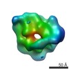 6055C 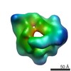 6056C 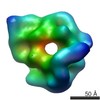 6058C 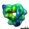 6059C  6060C  6061C  6062C  6063C  6064C 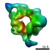 6065C 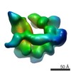 6066C 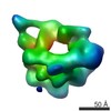 6067C 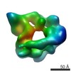 6068C  6069C  6070C  6071C  6072C 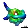 6073C 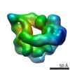 6074C C: citing same article ( |
|---|---|
| Similar structure data |

-Links
 PDBj
PDBj


- Assembly
| Deposited unit | 
| ||||||||
|---|---|---|---|---|---|---|---|---|---|
| 1 | 
| ||||||||
| 2 | 
| ||||||||
| 3 |
| ||||||||
| Unit cell |
|
-Components
| #1: Protein | Mass: 304889.875 Da / Num. of mol.: 2 / Mutation: E1849Q Source method: isolated from a genetically manipulated source Source: (gene. exp.) Saccharomyces cerevisiae (brewer's yeast), (gene. exp.) Enterobacteria phage T4 (virus)Strain: ATCC 204508 / S288c / Gene: DYN1, DHC1, YKR054C / Production host: Saccharomyces cerevisiae (brewer's yeast) / References: UniProt: P36022, UniProt: P00720, lysozyme#2: Chemical | ChemComp-ANP /   Mass: 506.196 Da / Num. of mol.: 8 / Source method: obtained synthetically / Formula: C10H17N6O12P3 / Comment: AMP-PNP, energy-carrying molecule analogue*YM Mass: 506.196 Da / Num. of mol.: 8 / Source method: obtained synthetically / Formula: C10H17N6O12P3 / Comment: AMP-PNP, energy-carrying molecule analogue*YM#3: Chemical |   Mass: 24.305 Da / Num. of mol.: 2 / Source method: obtained synthetically / Formula: Mg Mass: 24.305 Da / Num. of mol.: 2 / Source method: obtained synthetically / Formula: Mg |
|---|
-Experimental details
-Experiment
| Experiment | Method: X-RAY DIFFRACTION |
|---|
- Sample preparation
| Crystal | Density Matthews: 3.13 Å3/Da / Density % sol: 60.7 % / Description: thin plates |
|---|---|
| Crystal grow | Temperature: 295 K / Method: vapor diffusion, hanging drop / Details: 4-10% PEG 3350 and 200-300 mM NaAc |
-Data collection
| Diffraction | Mean temperature: 100 K |
|---|---|
| Diffraction source | Source: SYNCHROTRON / Site: ALS / Beamline: 8.3.1 / Wavelength: 1.1159 Å |
| Detector | Type: ADSC QUANTUM 315r / Detector: CCD / Date: Dec 6, 2012 |
| Radiation | Monochromator: Si 111 CHANNEL / Protocol: SINGLE WAVELENGTH / Monochromatic (M) / Laue (L): M / Scattering type: x-ray |
| Radiation wavelength | Wavelength: 1.1159 Å / Relative weight: 1 |
| Reflection | Resolution: 3.54→49.4 Å / Num. obs: 82983 / % possible obs: 92.2 % / Observed criterion σ(I): 3.3 / Redundancy: 10.9 % / Net I/σ(I): 8.9 |
- Processing
| Software | Name: PHENIX / Version: (phenix.refine: dev_1769) / Classification: refinement | |||||||||||||||||||||||||||||||||||||||||||||||||||||||||||||||||||||||||||||||||||||||||||||||||||||||||||||||||||||||||||||||||||||||||||||||||||||||||||||||||||||||||||||||||||||||||||||||||||||||||||||||||||||||||||||||||
|---|---|---|---|---|---|---|---|---|---|---|---|---|---|---|---|---|---|---|---|---|---|---|---|---|---|---|---|---|---|---|---|---|---|---|---|---|---|---|---|---|---|---|---|---|---|---|---|---|---|---|---|---|---|---|---|---|---|---|---|---|---|---|---|---|---|---|---|---|---|---|---|---|---|---|---|---|---|---|---|---|---|---|---|---|---|---|---|---|---|---|---|---|---|---|---|---|---|---|---|---|---|---|---|---|---|---|---|---|---|---|---|---|---|---|---|---|---|---|---|---|---|---|---|---|---|---|---|---|---|---|---|---|---|---|---|---|---|---|---|---|---|---|---|---|---|---|---|---|---|---|---|---|---|---|---|---|---|---|---|---|---|---|---|---|---|---|---|---|---|---|---|---|---|---|---|---|---|---|---|---|---|---|---|---|---|---|---|---|---|---|---|---|---|---|---|---|---|---|---|---|---|---|---|---|---|---|---|---|---|---|---|---|---|---|---|---|---|---|---|---|---|---|---|---|---|---|
| Refinement | Resolution: 3.541→49.4 Å / SU ML: 0.46 / Cross valid method: FREE R-VALUE / σ(F): 1.37 / Phase error: 25.86 / Stereochemistry target values: ML
| |||||||||||||||||||||||||||||||||||||||||||||||||||||||||||||||||||||||||||||||||||||||||||||||||||||||||||||||||||||||||||||||||||||||||||||||||||||||||||||||||||||||||||||||||||||||||||||||||||||||||||||||||||||||||||||||||
| Solvent computation | Shrinkage radii: 0.9 Å / VDW probe radii: 1.11 Å / Solvent model: FLAT BULK SOLVENT MODEL | |||||||||||||||||||||||||||||||||||||||||||||||||||||||||||||||||||||||||||||||||||||||||||||||||||||||||||||||||||||||||||||||||||||||||||||||||||||||||||||||||||||||||||||||||||||||||||||||||||||||||||||||||||||||||||||||||
| Refinement step | Cycle: LAST / Resolution: 3.541→49.4 Å
| |||||||||||||||||||||||||||||||||||||||||||||||||||||||||||||||||||||||||||||||||||||||||||||||||||||||||||||||||||||||||||||||||||||||||||||||||||||||||||||||||||||||||||||||||||||||||||||||||||||||||||||||||||||||||||||||||
| Refine LS restraints |
| |||||||||||||||||||||||||||||||||||||||||||||||||||||||||||||||||||||||||||||||||||||||||||||||||||||||||||||||||||||||||||||||||||||||||||||||||||||||||||||||||||||||||||||||||||||||||||||||||||||||||||||||||||||||||||||||||
| LS refinement shell |
| |||||||||||||||||||||||||||||||||||||||||||||||||||||||||||||||||||||||||||||||||||||||||||||||||||||||||||||||||||||||||||||||||||||||||||||||||||||||||||||||||||||||||||||||||||||||||||||||||||||||||||||||||||||||||||||||||
| Refinement TLS params. | Method: refined / Refine-ID: X-RAY DIFFRACTION
| |||||||||||||||||||||||||||||||||||||||||||||||||||||||||||||||||||||||||||||||||||||||||||||||||||||||||||||||||||||||||||||||||||||||||||||||||||||||||||||||||||||||||||||||||||||||||||||||||||||||||||||||||||||||||||||||||
| Refinement TLS group |
|
