 Movie
Movie Controller

[English] 日本語
 Yorodumi
- PDB-4w8c: Crystal structure of the helical domain deleted form MsrA from Cl... -
+ Open data

- Basic information
| Entry | Database: PDB / ID: 4w8c | ||||||
|---|---|---|---|---|---|---|---|
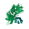
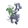
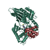
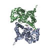
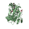
| Title | Crystal structure of the helical domain deleted form MsrA from Clostridium oremlandii | ||||||
 Components Components | Peptide methionine sulfoxide reductase MsrA | ||||||
 Keywords Keywords |  OXIDOREDUCTASE / MsrA / Clostridium oremlandii / truncated form OXIDOREDUCTASE / MsrA / Clostridium oremlandii / truncated form | ||||||
| Function / homology |  Function and homology informationL-methionine:thioredoxin-disulfide S-oxidoreductase activity / peptide-methionine (S)-S-oxide reductase / peptide-methionine (S)-S-oxide reductase activity / protein modification process Function and homology informationL-methionine:thioredoxin-disulfide S-oxidoreductase activity / peptide-methionine (S)-S-oxide reductase / peptide-methionine (S)-S-oxide reductase activity / protein modification processSimilarity search - Function | ||||||
| Biological species |  Alkaliphilus oremlandii OhILAs (bacteria) Alkaliphilus oremlandii OhILAs (bacteria) | ||||||
| Method | X-RAY DIFFRACTION / SYNCHROTRON / MOLECULAR REPLACEMENT / Resolution: 1.7568 Å | ||||||
 Authors Authors | Lee, E.H. / Hwang, K.Y. / Kim, H.-Y. | ||||||
 Citation Citation | Journal: Plos One / Year: 2015 Title: Essential role of the C-terminal helical domain in active site formation of selenoprotein MsrA from Clostridium oremlandii Authors: Lee, E.H. / Lee, K. / Hwang, K.Y. / Kim, H.-Y. | ||||||
| History |
|
- Structure visualization
| Structure viewer | Molecule: MolmilJmol/JSmol |
|---|
- Downloads & links
-Download
| PDBx/mmCIF format | 4w8c.cif.gz | 70.5 KB | Display | PDBx/mmCIF format |
|---|---|---|---|---|
| PDB format | pdb4w8c.ent.gz | 51.6 KB | Display | PDB format |
| PDBx/mmJSON format | 4w8c.json.gz | Tree view | PDBx/mmJSON format | |
| Others |  Other downloads Other downloads |
-Validation report
| Arichive directory | https://data.pdbj.org/pub/pdb/validation_reports/w8/4w8cftp://data.pdbj.org/pub/pdb/validation_reports/w8/4w8c | HTTPS FTP |
|---|
-Related structure data
| Related structure data | 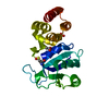 4lwjS S: Starting model for refinement |
|---|---|
| Similar structure data |


-Links
 PDBj
PDBj

- Assembly
| Deposited unit | 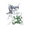
| |||||||||
|---|---|---|---|---|---|---|---|---|---|---|
| 1 |
| |||||||||
| Unit cell |
| |||||||||
| Components on special symmetry positions |
|
-Components
| #1: Protein | Mass: 16503.271 Da / Num. of mol.: 2 / Fragment: UNP residues 1-144 / Mutation: U16C Source method: isolated from a genetically manipulated source Source: (gene. exp.) Alkaliphilus oremlandii OhILAs (bacteria)Gene: msrA, Clos_1947 / Plasmid: pET21b / Production host: Escherichia coli (E. coli)References: UniProt: A8MI53, peptide-methionine (S)-S-oxide reductase#2: Chemical | Glycine  Type: peptide linking / Mass: 75.067 Da / Num. of mol.: 2 / Source method: obtained synthetically / Formula: C2H5NO2 Type: peptide linking / Mass: 75.067 Da / Num. of mol.: 2 / Source method: obtained synthetically / Formula: C2H5NO2#3: Water | ChemComp-HOH / | Water Mass: 18.015 Da / Num. of mol.: 115 / Source method: isolated from a natural source / Formula: H2O Mass: 18.015 Da / Num. of mol.: 115 / Source method: isolated from a natural source / Formula: H2O |
|---|
-Experimental details
-Experiment
| Experiment | Method: X-RAY DIFFRACTION / Number of used crystals: 1 |
|---|
- Sample preparation
| Crystal | Density Matthews: 3.04 Å3/Da / Density % sol: 59.58 % |
|---|---|
| Crystal grow | Temperature: 293 K / Method: vapor diffusion, hanging drop / pH: 8.5 / Details: 0.1 M Tris-HCl, 3.2 M NaCl |
-Data collection
| Diffraction | Mean temperature: 100 K |
|---|---|
| Diffraction source | Source: SYNCHROTRON / Site: PAL/PLS  / Beamline: 5C (4A) / Wavelength: 1 Å / Beamline: 5C (4A) / Wavelength: 1 Å |
| Detector | Type: ADSC QUANTUM 315r / Detector: CCD / Date: Nov 19, 2013 |
| Radiation | Protocol: SINGLE WAVELENGTH / Monochromatic (M) / Laue (L): M / Scattering type: x-ray |
| Radiation wavelength | Wavelength: 1 Å / Relative weight: 1 |
| Reflection | Resolution: 1.7→50 Å / Num. obs: 36941 / % possible obs: 96.4 % / Redundancy: 8.9 % / Biso Wilson estimate: 26.65 Å2 / Net I/σ(I): 49.7 |
- Processing
| Software |
| |||||||||||||||||||||||||||||||||||||||||||||||||||||||||||||||||||||||||||||||||||||||||||||||||||||||||
|---|---|---|---|---|---|---|---|---|---|---|---|---|---|---|---|---|---|---|---|---|---|---|---|---|---|---|---|---|---|---|---|---|---|---|---|---|---|---|---|---|---|---|---|---|---|---|---|---|---|---|---|---|---|---|---|---|---|---|---|---|---|---|---|---|---|---|---|---|---|---|---|---|---|---|---|---|---|---|---|---|---|---|---|---|---|---|---|---|---|---|---|---|---|---|---|---|---|---|---|---|---|---|---|---|---|---|
| Refinement | Method to determine structure: MOLECULAR REPLACEMENT Starting model: 4LWJ Resolution: 1.7568→32.6 Å / FOM work R set: 0.7456 / SU ML: 0.25 / Cross valid method: FREE R-VALUE / σ(F): 0.11 / Phase error: 31.49 / Stereochemistry target values: ML
| |||||||||||||||||||||||||||||||||||||||||||||||||||||||||||||||||||||||||||||||||||||||||||||||||||||||||
| Solvent computation | Shrinkage radii: 0.9 Å / VDW probe radii: 1.11 Å / Solvent model: FLAT BULK SOLVENT MODEL | |||||||||||||||||||||||||||||||||||||||||||||||||||||||||||||||||||||||||||||||||||||||||||||||||||||||||
| Displacement parameters | Biso max: 84.77 Å2 / Biso mean: 33.92 Å2 / Biso min: 11.13 Å2 | |||||||||||||||||||||||||||||||||||||||||||||||||||||||||||||||||||||||||||||||||||||||||||||||||||||||||
| Refinement step | Cycle: final / Resolution: 1.7568→32.6 Å
| |||||||||||||||||||||||||||||||||||||||||||||||||||||||||||||||||||||||||||||||||||||||||||||||||||||||||
| Refine LS restraints |
| |||||||||||||||||||||||||||||||||||||||||||||||||||||||||||||||||||||||||||||||||||||||||||||||||||||||||
| LS refinement shell | Refine-ID: X-RAY DIFFRACTION / Total num. of bins used: 14
|
