 Movie
Movie Controller

[English] 日本語
 Yorodumi
- PDB-2p7i: CRYSTAL STRUCTURE OF a SAM dependent methyl-transferase type 12 f... -
+ Open data

- Basic information
| Entry | Database: PDB / ID: 2p7i | ||||||
|---|---|---|---|---|---|---|---|
| Title | CRYSTAL STRUCTURE OF a SAM dependent methyl-transferase type 12 family protein (ECA1738) FROM PECTOBACTERIUM ATROSEPTICUM SCRI1043 AT 1.74 A RESOLUTION | ||||||
 Components Components | Hypothetical protein | ||||||
 Keywords Keywords | TRANSFERASE / PUTATIVE METHYLTRANSFERASE / STRUCTURAL GENOMICS / JOINT CENTER FOR STRUCTURAL GENOMICS / JCSG / PROTEIN STRUCTURE INITIATIVE / PSI-2 | ||||||
| Function / homology | Methyltransferase domain / Vaccinia Virus protein VP39 / transferase activity / S-adenosyl-L-methionine-dependent methyltransferase superfamily / Rossmann fold / 3-Layer(aba) Sandwich / Alpha Beta / Methyltransferase Function and homology information Function and homology information | ||||||
| Biological species |  Pectobacterium atrosepticum SCRI1043 (bacteria) Pectobacterium atrosepticum SCRI1043 (bacteria) | ||||||
| Method | X-RAY DIFFRACTION / SYNCHROTRON / MAD / Resolution: 1.74 Å | ||||||
 Authors Authors | Joint Center for Structural Genomics (JCSG) | ||||||
 Citation Citation | Journal: To be published Title: Crystal structure of hypothetical protein (YP_049838.1) from Erwinia carotovora atroseptica SCRI1043 at 1.74 A resolution Authors: Joint Center for Structural Genomics (JCSG) | ||||||
| History |
| ||||||
| Remark 999 | SEQUENCE THE CONSTRUCT WAS EXPRESSED WITH A PURIFICATION TAG MGSDKIHHHHHHENLYFQG. THE TAG WAS ... SEQUENCE THE CONSTRUCT WAS EXPRESSED WITH A PURIFICATION TAG MGSDKIHHHHHHENLYFQG. THE TAG WAS REMOVED WITH TEV PROTEASE LEAVING ONLY A GLYCINE, FOLLOWED BY THE TARGET SEQUENCE. |
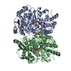
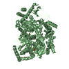
- Structure visualization
| Structure viewer | Molecule: MolmilJmol/JSmol |
|---|
- Downloads & links
-Download
| PDBx/mmCIF format | 2p7i.cif.gz | 113.3 KB | Display | PDBx/mmCIF format |
|---|---|---|---|---|
| PDB format | pdb2p7i.ent.gz | 90.7 KB | Display | PDB format |
| PDBx/mmJSON format | 2p7i.json.gz | Tree view | PDBx/mmJSON format | |
| Others |  Other downloads Other downloads |
-Validation report
| Arichive directory | https://data.pdbj.org/pub/pdb/validation_reports/p7/2p7iftp://data.pdbj.org/pub/pdb/validation_reports/p7/2p7i | HTTPS FTP |
|---|
-Related structure data
| Related structure data | |
|---|---|
| Similar structure data | |
| Other databases |


-Links
 PDBj
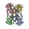
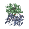
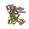
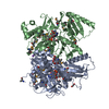
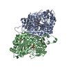
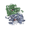
PDBj- Assembly
| Deposited unit | 
| |||||||||||||||||||||
|---|---|---|---|---|---|---|---|---|---|---|---|---|---|---|---|---|---|---|---|---|---|---|
| 1 | 
| |||||||||||||||||||||
| 2 | x 8
| |||||||||||||||||||||
| Unit cell |
| |||||||||||||||||||||
| Components on special symmetry positions |
| |||||||||||||||||||||
| Noncrystallographic symmetry (NCS) | NCS domain: (Details: A B) NCS domain segments: Dom-ID: 1 / Ens-ID: 1 / Refine code: 4
|
-Components
-Protein , 1 types, 2 molecules AB
| #1: Protein | Mass: 28685.703 Da / Num. of mol.: 2 Source method: isolated from a genetically manipulated source Source: (gene. exp.) Pectobacterium atrosepticum SCRI1043 (bacteria)Species: Pectobacterium atrosepticum / Strain: SCRI 1043 / Gene: YP_049838.1, ECA1738 / Plasmid: speedET / Production host: Escherichia coli (E. coli) / Strain (production host): HK100 / References: UniProt: Q6D6E7 |
|---|
-Non-polymers , 5 types, 371 molecules 


| #2: Chemical | ChemComp-CL / Chloride Mass: 35.453 Da / Num. of mol.: 1 / Source method: obtained synthetically / Formula: Cl Mass: 35.453 Da / Num. of mol.: 1 / Source method: obtained synthetically / Formula: Cl | ||||||
|---|---|---|---|---|---|---|---|
| #3: Chemical | Tris Mass: 122.143 Da / Num. of mol.: 2 / Source method: obtained synthetically / Formula: C4H12NO3 / Comment: pH buffer*YM Mass: 122.143 Da / Num. of mol.: 2 / Source method: obtained synthetically / Formula: C4H12NO3 / Comment: pH buffer*YM#4: Chemical | ChemComp-MPD / ( | 2-Methyl-2,4-pentanediol Mass: 118.174 Da / Num. of mol.: 1 / Source method: obtained synthetically / Formula: C6H14O2 / Comment: precipitant*YM Mass: 118.174 Da / Num. of mol.: 1 / Source method: obtained synthetically / Formula: C6H14O2 / Comment: precipitant*YM#5: Chemical | ChemComp-NA / |  Mass: 22.990 Da / Num. of mol.: 1 / Source method: obtained synthetically / Formula: Na Mass: 22.990 Da / Num. of mol.: 1 / Source method: obtained synthetically / Formula: Na#6: Water | ChemComp-HOH / | WaterMass: 18.015 Da / Num. of mol.: 366 / Source method: isolated from a natural source / Formula: H2O |
-Experimental details
-Experiment
| Experiment | Method: X-RAY DIFFRACTION / Number of used crystals: 1 |
|---|
- Sample preparation
| Crystal | Density Matthews: 2.38 Å3/Da / Density % sol: 48.42 % |
|---|---|
| Crystal grow | Temperature: 277 K / Method: vapor diffusion, sitting drop / pH: 7.5 Details: NANODROP, 64.4% 2-methyl-2,4-pentanediol, 0.1M Tris-HCl pH 7.5, VAPOR DIFFUSION, SITTING DROP, temperature 277K |
-Data collection
| Diffraction | Mean temperature: 100 K | |||||||||||||||||||||||||||||||||||||||||||||||||||||||||||||||||||||||||||||||||||||||||||||||||||||||||||||||||||||||||||||||||||||||||||||||||||
|---|---|---|---|---|---|---|---|---|---|---|---|---|---|---|---|---|---|---|---|---|---|---|---|---|---|---|---|---|---|---|---|---|---|---|---|---|---|---|---|---|---|---|---|---|---|---|---|---|---|---|---|---|---|---|---|---|---|---|---|---|---|---|---|---|---|---|---|---|---|---|---|---|---|---|---|---|---|---|---|---|---|---|---|---|---|---|---|---|---|---|---|---|---|---|---|---|---|---|---|---|---|---|---|---|---|---|---|---|---|---|---|---|---|---|---|---|---|---|---|---|---|---|---|---|---|---|---|---|---|---|---|---|---|---|---|---|---|---|---|---|---|---|---|---|---|---|---|---|
| Diffraction source | Source: SYNCHROTRON / Site: SSRL  / Beamline: BL11-1 / Wavelength: 0.97899, 0.97925 / Beamline: BL11-1 / Wavelength: 0.97899, 0.97925 | |||||||||||||||||||||||||||||||||||||||||||||||||||||||||||||||||||||||||||||||||||||||||||||||||||||||||||||||||||||||||||||||||||||||||||||||||||
| Detector | Type: MARMOSAIC 325 mm CCD / Detector: CCD / Date: Mar 2, 2007 / Details: Flat mirror (vertical focusing) | |||||||||||||||||||||||||||||||||||||||||||||||||||||||||||||||||||||||||||||||||||||||||||||||||||||||||||||||||||||||||||||||||||||||||||||||||||
| Radiation | Monochromator: Single crystal Si(111) bent (horizontal focusing) Protocol: MAD / Monochromatic (M) / Laue (L): M / Scattering type: x-ray | |||||||||||||||||||||||||||||||||||||||||||||||||||||||||||||||||||||||||||||||||||||||||||||||||||||||||||||||||||||||||||||||||||||||||||||||||||
| Radiation wavelength |
| |||||||||||||||||||||||||||||||||||||||||||||||||||||||||||||||||||||||||||||||||||||||||||||||||||||||||||||||||||||||||||||||||||||||||||||||||||
| Reflection | Resolution: 1.74→29.111 Å / Num. obs: 56875 / % possible obs: 100 % / Redundancy: 7.3 % / Biso Wilson estimate: 25.14 Å2 / Rmerge(I) obs: 0.09 / Rsym value: 0.09 / Net I/σ(I): 6.3 | |||||||||||||||||||||||||||||||||||||||||||||||||||||||||||||||||||||||||||||||||||||||||||||||||||||||||||||||||||||||||||||||||||||||||||||||||||
| Reflection shell | Rmerge(I) obs: 0.011 / Diffraction-ID: 1
|
-Phasing
| Phasing | Method: MAD |
|---|
- Processing
| Software |
| |||||||||||||||||||||||||||||||||||||||||||||||||||||||||||||||||||||||||||||||||||||||||||||||||||||||||||||||||||||||||||||||||||||||
|---|---|---|---|---|---|---|---|---|---|---|---|---|---|---|---|---|---|---|---|---|---|---|---|---|---|---|---|---|---|---|---|---|---|---|---|---|---|---|---|---|---|---|---|---|---|---|---|---|---|---|---|---|---|---|---|---|---|---|---|---|---|---|---|---|---|---|---|---|---|---|---|---|---|---|---|---|---|---|---|---|---|---|---|---|---|---|---|---|---|---|---|---|---|---|---|---|---|---|---|---|---|---|---|---|---|---|---|---|---|---|---|---|---|---|---|---|---|---|---|---|---|---|---|---|---|---|---|---|---|---|---|---|---|---|---|---|
| Refinement | Method to determine structure: MAD / Resolution: 1.74→29.111 Å / Cor.coef. Fo:Fc: 0.976 / Cor.coef. Fo:Fc free: 0.968 / SU B: 4.556 / SU ML: 0.073 / TLS residual ADP flag: LIKELY RESIDUAL / Cross valid method: THROUGHOUT / σ(F): 0 / ESU R: 0.091 / ESU R Free: 0.088 Stereochemistry target values: MAXIMUM LIKELIHOOD WITH PHASES Details: 1. HYDROGENS HAVE BEEN ADDED IN THE RIDING POSITIONS. 2. ATOM RECORD CONTAINS RESIDUAL B FACTORS ONLY. 3. A MET-INHIBITION PROTOCOL WAS USED FOR SELENOMETHIONINE INCORPORATION DURING PROTEIN ...Details: 1. HYDROGENS HAVE BEEN ADDED IN THE RIDING POSITIONS. 2. ATOM RECORD CONTAINS RESIDUAL B FACTORS ONLY. 3. A MET-INHIBITION PROTOCOL WAS USED FOR SELENOMETHIONINE INCORPORATION DURING PROTEIN EXPRESSION. THE OCCUPANCY OF THE SE ATOMS IN THE MSE RESIDUES WAS REDUCED TO 0.75 FOR THE REDUCED SCATTERING POWER DUE TO PARTIAL S-MET INCORPORATION. 4. TRS, CL, NA AND MPD ARE MODELED BASED ON CRYSTALLIZATION CONDITIONS. 5. THE N-TERMINAL RESIDUES 0-21 FOR CHAIN A, AND 0-20 FOR CHAIN B ARE DISORDERED. 6. RAMACHANDRAN OUTLIERS FOR RESIDUE 199 ARE SUPPORTED BY UNAMBIGUOUS ELECTRON DENSITY.
| |||||||||||||||||||||||||||||||||||||||||||||||||||||||||||||||||||||||||||||||||||||||||||||||||||||||||||||||||||||||||||||||||||||||
| Solvent computation | Ion probe radii: 0.8 Å / Shrinkage radii: 0.8 Å / VDW probe radii: 1.2 Å / Solvent model: BABINET MODEL WITH MASK | |||||||||||||||||||||||||||||||||||||||||||||||||||||||||||||||||||||||||||||||||||||||||||||||||||||||||||||||||||||||||||||||||||||||
| Displacement parameters | Biso mean: 26.628 Å2
| |||||||||||||||||||||||||||||||||||||||||||||||||||||||||||||||||||||||||||||||||||||||||||||||||||||||||||||||||||||||||||||||||||||||
| Refinement step | Cycle: LAST / Resolution: 1.74→29.111 Å
| |||||||||||||||||||||||||||||||||||||||||||||||||||||||||||||||||||||||||||||||||||||||||||||||||||||||||||||||||||||||||||||||||||||||
| Refine LS restraints |
| |||||||||||||||||||||||||||||||||||||||||||||||||||||||||||||||||||||||||||||||||||||||||||||||||||||||||||||||||||||||||||||||||||||||
| Refine LS restraints NCS | Dom-ID: 1 / Ens-ID: 1 / Number: 2916 / Refine-ID: X-RAY DIFFRACTION
| |||||||||||||||||||||||||||||||||||||||||||||||||||||||||||||||||||||||||||||||||||||||||||||||||||||||||||||||||||||||||||||||||||||||
| LS refinement shell | Resolution: 1.74→1.785 Å / Total num. of bins used: 20
| |||||||||||||||||||||||||||||||||||||||||||||||||||||||||||||||||||||||||||||||||||||||||||||||||||||||||||||||||||||||||||||||||||||||
| Refinement TLS params. | Method: refined / Refine-ID: X-RAY DIFFRACTION
| |||||||||||||||||||||||||||||||||||||||||||||||||||||||||||||||||||||||||||||||||||||||||||||||||||||||||||||||||||||||||||||||||||||||
| Refinement TLS group | Refine-ID: X-RAY DIFFRACTION / Selection: ALL
|
