 Movie
Movie Controller

[English] 日本語
 Yorodumi
- PDB-4uyk: Crystal structure of a Signal Recognition Particle Alu domain in ... -
+ Open data

- Basic information
| Entry | Database: PDB / ID: 4uyk | ||||||
|---|---|---|---|---|---|---|---|
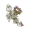
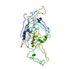
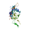
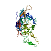
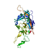
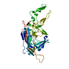
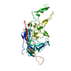
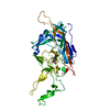
| Title | Crystal structure of a Signal Recognition Particle Alu domain in the elongation arrest conformation | ||||||
 Components Components |
| ||||||
 Keywords Keywords |  SIGNALING PROTEIN / SIGNAL RECOGNITION PARTICLE / TRANSLATION / RNA / RNA FOLDING SIGNALING PROTEIN / SIGNAL RECOGNITION PARTICLE / TRANSLATION / RNA / RNA FOLDING | ||||||
| Function / homology |  Function and homology informationsignal recognition particle receptor complex / endoplasmic reticulum signal peptide binding / signal recognition particle, endoplasmic reticulum targeting / signal recognition particle binding / negative regulation of translational elongation / cotranslational protein targeting to membrane / protein targeting to ER / 7S RNA binding / SRP-dependent cotranslational protein targeting to membrane / SRP-dependent cotranslational protein targeting to membrane ...signal recognition particle receptor complex / endoplasmic reticulum signal peptide binding / signal recognition particle, endoplasmic reticulum targeting / signal recognition particle binding / negative regulation of translational elongation / cotranslational protein targeting to membrane / protein targeting to ER / 7S RNA binding / SRP-dependent cotranslational protein targeting to membrane / SRP-dependent cotranslational protein targeting to membrane / secretory granule lumen / ficolin-1-rich granule lumen / Neutrophil degranulation / RNA binding / extracellular region / nucleus / cytosol / cytoplasm Function and homology informationsignal recognition particle receptor complex / endoplasmic reticulum signal peptide binding / signal recognition particle, endoplasmic reticulum targeting / signal recognition particle binding / negative regulation of translational elongation / cotranslational protein targeting to membrane / protein targeting to ER / 7S RNA binding / SRP-dependent cotranslational protein targeting to membrane / SRP-dependent cotranslational protein targeting to membrane ...signal recognition particle receptor complex / endoplasmic reticulum signal peptide binding / signal recognition particle, endoplasmic reticulum targeting / signal recognition particle binding / negative regulation of translational elongation / cotranslational protein targeting to membrane / protein targeting to ER / 7S RNA binding / SRP-dependent cotranslational protein targeting to membrane / SRP-dependent cotranslational protein targeting to membrane / secretory granule lumen / ficolin-1-rich granule lumen / Neutrophil degranulation / RNA binding / extracellular region / nucleus / cytosol / cytoplasmSimilarity search - Function | ||||||
| Biological species |  HOMO SAPIENS (human) HOMO SAPIENS (human)  PYROCOCCUS HORIKOSHII OT3 (archaea) PYROCOCCUS HORIKOSHII OT3 (archaea) | ||||||
| Method | X-RAY DIFFRACTION / SYNCHROTRON / MAD / Resolution: 3.22 Å | ||||||
 Authors Authors | Bousset, L. / Mary, C. / Brooks, M.A. / Scherrer, A. / Strub, K. / Cusack, S. | ||||||
 Citation Citation | Journal: RNA / Year: 2014 Title: Crystal Structure of a Signal Recognition Particle Alu Domain in the Elongation Arrest Conformation. Authors: Bousset, L. / Mary, C. / Brooks, M.A. / Scherrer, A. / Strub, K. / Cusack, S. | ||||||
| History |
|
- Structure visualization
| Structure viewer | Molecule: MolmilJmol/JSmol |
|---|
- Downloads & links
-Download
| PDBx/mmCIF format | 4uyk.cif.gz | 226.3 KB | Display | PDBx/mmCIF format |
|---|---|---|---|---|
| PDB format | pdb4uyk.ent.gz | 190.1 KB | Display | PDB format |
| PDBx/mmJSON format | 4uyk.json.gz | Tree view | PDBx/mmJSON format | |
| Others |  Other downloads Other downloads |
-Validation report
| Arichive directory | https://data.pdbj.org/pub/pdb/validation_reports/uy/4uykftp://data.pdbj.org/pub/pdb/validation_reports/uy/4uyk | HTTPS FTP |
|---|
-Related structure data


-Links
 PDBj
PDBj


- Assembly
| Deposited unit | 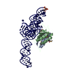
| ||||||||
|---|---|---|---|---|---|---|---|---|---|
| 1 |
| ||||||||
| Unit cell |
|
-Components
| #1: Protein | Mass: 10233.126 Da / Num. of mol.: 1 / Fragment: RESIDUES 1-85 Source method: isolated from a genetically manipulated source Source: (gene. exp.) HOMO SAPIENS (human) / Plasmid: PEH9 / Production host:  ESCHERICHIA COLI (E. coli) / Strain (production host): BL21(DE3) / References: UniProt: P49458 ESCHERICHIA COLI (E. coli) / Strain (production host): BL21(DE3) / References: UniProt: P49458 |
|---|---|
| #2: Protein | Mass: 12386.117 Da / Num. of mol.: 1 / Fragment: RESIDUES 1-107 Source method: isolated from a genetically manipulated source Source: (gene. exp.) HOMO SAPIENS (human) / Plasmid: PEH14DELTAR / Production host: ESCHERICHIA COLI (E. coli) / Strain (production host): BL21(DE3) / References: UniProt: P37108 |
| #3: RNA chain | Signal recognition particle RNA Mass: 43523.797 Da / Num. of mol.: 1 / Fragment: ALU DOMAIN, RESIDUES 1-89 AND RESIDUES 289-314 / Source method: obtained synthetically / Source: (synth.) PYROCOCCUS HORIKOSHII OT3 (archaea) / References: GenBank: HG323574 |
| Sequence details | HUMAN SRP9 RESIDUES 1 TO 85 WERE EXPRESSED IN THIS WORK HUMAN SRP14 RESIDUES 1 TO 107 WERE ...HUMAN SRP9 RESIDUES 1 TO 85 WERE EXPRESSED IN THIS WORK HUMAN SRP14 RESIDUES 1 TO 107 WERE EXPRESSED IN THIS WORK THE P. HORIKOSHII |
-Experimental details
-Experiment
| Experiment | Method: X-RAY DIFFRACTION / Number of used crystals: 1 |
|---|
- Sample preparation
| Crystal | Density Matthews: 4.12 Å3/Da / Density % sol: 73.3 % / Description: NONE |
|---|---|
| Crystal grow | pH: 5 Details: 17.5% PEG 400, 5% GLYCEROL BUFFERED IN 100 MM SODIUM ACETATE PH 5.0 |
-Data collection
| Diffraction | Mean temperature: 100 K |
|---|---|
| Diffraction source | Source: SYNCHROTRON / Site: ESRF  / Beamline: ID14-1 / Wavelength: 0.934 / Beamline: ID14-1 / Wavelength: 0.934 |
| Detector | Type: ADSC CCD / Detector: CCD / Date: Apr 16, 2004 |
| Radiation | Monochromator: DIAMOND (111) / Protocol: MAD / Monochromatic (M) / Laue (L): M / Scattering type: x-ray |
| Radiation wavelength | Wavelength: 0.934 Å / Relative weight: 1 |
| Reflection | Resolution: 3.2→30 Å / Num. obs: 30821 / % possible obs: 97.8 % / Observed criterion σ(I): 0 / Redundancy: 2.18 % / Rmerge(I) obs: 0.08 / Net I/σ(I): 9.09 |
| Reflection shell | Resolution: 3.2→3.4 Å / Redundancy: 2.18 % / Rmerge(I) obs: 0.64 / Mean I/σ(I) obs: 1.74 / % possible all: 95.9 |
- Processing
| Software |
| ||||||||||||||||||||||||||||||||||||||||||||||||||||||||||||||||||||||||||||||||||||||||||||||||||||||||||||||||||||||||||||||||||||||||||||||||||||||||||||||||||||||||||||||||||||||
|---|---|---|---|---|---|---|---|---|---|---|---|---|---|---|---|---|---|---|---|---|---|---|---|---|---|---|---|---|---|---|---|---|---|---|---|---|---|---|---|---|---|---|---|---|---|---|---|---|---|---|---|---|---|---|---|---|---|---|---|---|---|---|---|---|---|---|---|---|---|---|---|---|---|---|---|---|---|---|---|---|---|---|---|---|---|---|---|---|---|---|---|---|---|---|---|---|---|---|---|---|---|---|---|---|---|---|---|---|---|---|---|---|---|---|---|---|---|---|---|---|---|---|---|---|---|---|---|---|---|---|---|---|---|---|---|---|---|---|---|---|---|---|---|---|---|---|---|---|---|---|---|---|---|---|---|---|---|---|---|---|---|---|---|---|---|---|---|---|---|---|---|---|---|---|---|---|---|---|---|---|---|---|---|
| Refinement | Method to determine structure: MAD Starting model: NONE Resolution: 3.22→89.82 Å / Cor.coef. Fo:Fc: 0.957 / Cor.coef. Fo:Fc free: 0.936 / SU B: 48.231 / SU ML: 0.338 / Cross valid method: THROUGHOUT / ESU R Free: 0.392 / Stereochemistry target values: MAXIMUM LIKELIHOOD Details: HYDROGENS HAVE BEEN ADDED IN THE RIDING POSITIONS. U VALUES WITH TLS ADDED
| ||||||||||||||||||||||||||||||||||||||||||||||||||||||||||||||||||||||||||||||||||||||||||||||||||||||||||||||||||||||||||||||||||||||||||||||||||||||||||||||||||||||||||||||||||||||
| Solvent computation | Ion probe radii: 0.8 Å / Shrinkage radii: 0.8 Å / VDW probe radii: 1.2 Å / Solvent model: MASK | ||||||||||||||||||||||||||||||||||||||||||||||||||||||||||||||||||||||||||||||||||||||||||||||||||||||||||||||||||||||||||||||||||||||||||||||||||||||||||||||||||||||||||||||||||||||
| Displacement parameters | Biso mean: 123.705 Å2
| ||||||||||||||||||||||||||||||||||||||||||||||||||||||||||||||||||||||||||||||||||||||||||||||||||||||||||||||||||||||||||||||||||||||||||||||||||||||||||||||||||||||||||||||||||||||
| Refinement step | Cycle: LAST / Resolution: 3.22→89.82 Å
| ||||||||||||||||||||||||||||||||||||||||||||||||||||||||||||||||||||||||||||||||||||||||||||||||||||||||||||||||||||||||||||||||||||||||||||||||||||||||||||||||||||||||||||||||||||||
| Refine LS restraints |
|