Movie
Movie Controller

[English] 日本語
 Yorodumi
- PDB-3j1q: Structure of AAV-DJ, a Retargeted Gene Therapy Vector: Cryo-Elect... -
+ Open data

- Basic information
| Entry | Database: PDB / ID: 3j1q | ||||||
|---|---|---|---|---|---|---|---|
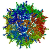
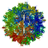
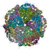
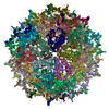
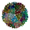
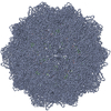
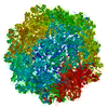
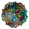
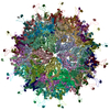
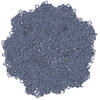
| Title | Structure of AAV-DJ, a Retargeted Gene Therapy Vector: Cryo-Electron Microscopy at 4.5A resolution | ||||||
 Components Components | Adeno-associated virus DJ | ||||||
 Keywords Keywords |  VIRUS / Gene therapy VIRUS / Gene therapy | ||||||
| Function / homology | Phospholipase A2-like domain / Phospholipase A2-like domain / Parvovirus coat protein VP2 / Parvovirus coat protein VP1/VP2 / Parvovirus coat protein VP2 / Capsid/spike protein, ssDNA virus / T=1 icosahedral viral capsid / structural molecule activity / Capsid protein VP1 Function and homology information Function and homology information | ||||||
| Biological species |   Adeno-associated virus Adeno-associated virus | ||||||
| Method | ELECTRON MICROSCOPY / single particle reconstruction / cryo EM / Resolution: 4.5 Å | ||||||
 Authors Authors | Lerch, T.F. / O'Donnell, J.K. / Meyer, N.L. / Xie, Q. / Taylor, K.A. / Stagg, S.M. / Chapman, M.S. | ||||||
 Citation Citation | Journal: Structure / Year: 2012 Title: Structure of AAV-DJ, a retargeted gene therapy vector: cryo-electron microscopy at 4.5 Å resolution. Authors: Thomas F Lerch / Jason K O'Donnell / Nancy L Meyer / Qing Xie / Kenneth A Taylor / Scott M Stagg / Michael S Chapman /  Abstract: AAV-DJ, a leading candidate vector for liver gene therapy, was created through random homologous recombination followed by directed evolution, selecting for in vivo liver tropism and resistance to ...AAV-DJ, a leading candidate vector for liver gene therapy, was created through random homologous recombination followed by directed evolution, selecting for in vivo liver tropism and resistance to in vitro immune neutralization. Here, the 4.5 Å resolution cryo-EM structure is determined for the engineered AAV vector, revealing structural features that illuminate its phenotype. The heparan sulfate receptor-binding site is little changed from AAV-2, and heparin-binding affinity is similar. A loop that is antigenic in other serotypes has a unique conformation in AAV-DJ that would conflict with the binding of an AAV-2 neutralizing monoclonal antibody. This is consistent with increased resistance to neutralization by human polyclonal sera, raising the possibility that changed tropism may be a secondary effect of altered immune interactions. The reconstruction exemplifies analysis of fine structural changes and the potential of cryo-EM, in favorable cases, to characterize mutant or ligand-bound complexes. | ||||||
| History |
|
- Structure visualization
| Movie |
Movie viewer |
|---|---|
| Structure viewer | Molecule: MolmilJmol/JSmol |
- Downloads & links
-Download
| PDBx/mmCIF format | 3j1q.cif.gz | 117.8 KB | Display | PDBx/mmCIF format |
|---|---|---|---|---|
| PDB format | pdb3j1q.ent.gz | 88.5 KB | Display | PDB format |
| PDBx/mmJSON format | 3j1q.json.gz | Tree view | PDBx/mmJSON format | |
| Others |  Other downloads Other downloads |
-Validation report
| Arichive directory | https://data.pdbj.org/pub/pdb/validation_reports/j1/3j1qftp://data.pdbj.org/pub/pdb/validation_reports/j1/3j1q | HTTPS FTP |
|---|
-Related structure data
| Related structure data | 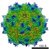 5415MC M: map data used to model this data C: citing same article ( |
|---|---|
| Similar structure data |


-Links
 PDBj
PDBj


- Assembly
| Deposited unit | 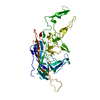
|
|---|---|
| 1 | x 60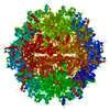
|
| 2 |
|
| 3 | x 5
|
| 4 | x 6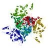
|
| 5 | 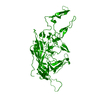
|
| Symmetry | Point symmetry: (Schoenflies symbol: I (icosahedral)) |
-Components
| #1: Protein | Mass: 82008.406 Da / Num. of mol.: 1 Source method: isolated from a genetically manipulated source Source: (gene. exp.) Adeno-associated virus / Strain: hybrid of serotypes 2, 8 and 9 / Gene: Capsid / Plasmid: pFBDDJm11 / Production host:   Spodoptera frugiperda (fall armyworm) / Strain (production host): SF9 / References: UniProt: Q6JC41*PLUS Spodoptera frugiperda (fall armyworm) / Strain (production host): SF9 / References: UniProt: Q6JC41*PLUS |
|---|
-Experimental details
-Experiment
| Experiment | Method: ELECTRON MICROSCOPY |
|---|---|
| EM experiment | Aggregation state: PARTICLE / 3D reconstruction method: single particle reconstruction |
- Sample preparation
| Component | Name: Adeno-associated virus DJ / Type: COMPLEX Details: 60 subunits associate to form a T=1 icosahedral capsid |
|---|---|
| Details of virus | Host category: VERTEBRATES / Isolate: SEROTYPE / Type: VIRUS-LIKE PARTICLE |
| Natural host | Organism: Homo sapiens |
| Buffer solution | Name: 125 mM sodium chloride, 10 mM Tris, 1 mM magnesium chloride pH: 7.5 Details: 125 mM sodium chloride, 10 mM Tris, 1 mM magnesium chloride |
| Specimen | Conc.: 1.5 mg/ml / Embedding applied: NO / Shadowing applied: NO / Staining applied: NO / Vitrification applied: YES |
| Specimen support | Details: Sample prepared on C-flat 2um hole, 1 um spacing, 200 mesh copper grids |
| Vitrification | Instrument: FEI VITROBOT MARK I / Cryogen name: ETHANE |
- Electron microscopy imaging
| Experimental equipment |  Model: Titan Krios / Image courtesy: FEI Company |
|---|---|
| Microscopy | Model: FEI TITAN KRIOS / Date: Feb 25, 2011 |
| Electron gun | Electron source: FIELD EMISSION GUN / Accelerating voltage: 300 kV / Illumination mode: FLOOD BEAM |
| Electron lens | Mode: BRIGHT FIELDBright-field microscopy |
| Specimen holder | Specimen holder model: OTHER |
| Image scans | Num. digital images: 4773 |
| Radiation | Protocol: SINGLE WAVELENGTH / Monochromatic (M) / Laue (L): M / Scattering type: x-ray |
| Radiation wavelength | Relative weight: 1 |
- Processing
| EM software |
| ||||||||||||||||
|---|---|---|---|---|---|---|---|---|---|---|---|---|---|---|---|---|---|
| CTF correction | Details: CTF was estimated using Appion software | ||||||||||||||||
| Symmetry | Point symmetry: I (icosahedral) | ||||||||||||||||
| 3D reconstruction | Method: single particle, icosahedralSingle particle analysis Resolution: 4.5 Å / Resolution method: FSC 0.143 CUT-OFF / Num. of particles: 27312 / Nominal pixel size: 1.3217 Å / Actual pixel size: 1.3217 Å Magnification calibration: Magnification was calibrated against the crystal structure of a known homolog, AAV2 (1LP3) Details: Frealign was used for particle refinement in the reconstruction Symmetry type: POINT | ||||||||||||||||
| Atomic model building | B value: 30 / Protocol: OTHER / Space: REAL / Target criteria: Cross-correlation coefficient Details: REFINEMENT PROTOCOL--simulated annealing torsion angle dynamics, isotropic B-factor refinement DETAILS--Iterative refinement in RSRef and model building in Coot | ||||||||||||||||
| Atomic model building | PDB-ID: 1LP3 Pdb chain-ID: A / Accession code: 1LP3 / Source name: PDB / Type: experimental model | ||||||||||||||||
| Refinement step | Cycle: LAST
|