 Movie
Movie Controller

[English] 日本語
 Yorodumi
- PDB-4uvr: Binding mode, selectivity and potency of N-indolyl-oxopyridinyl-4... -
+ Open data

- Basic information
| Entry | Database: PDB / ID: 4uvr | ||||||
|---|---|---|---|---|---|---|---|
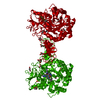
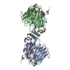
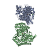
| Title | Binding mode, selectivity and potency of N-indolyl-oxopyridinyl-4- amino-propanyl-based inhibitors targeting Trypanosoma cruzi CYP51 | ||||||
 Components Components | STEROL 14-DEMETHYLASE, CYP51 | ||||||
 Keywords Keywords | OXIDOREDUCTASE / CYP51 / STEROL 14-DEMETHYLASE / STEROL BIOSYNTHESIS / CHAGAS DISEASE | ||||||
| Function / homology |  Function and homology information Function and homology informationsterol 14alpha-demethylase / sterol 14-demethylase activity / steroid 7-alpha-hydroxylase activity / oxysterol 7-alpha-hydroxylase activity / sterol biosynthetic process / bile acid biosynthetic process / cholesterol homeostasis / iron ion binding / heme binding / membraneSimilarity search - Function | ||||||
| Biological species |  TRYPANOSOMA CRUZI (eukaryote) TRYPANOSOMA CRUZI (eukaryote) | ||||||
| Method | X-RAY DIFFRACTION / SYNCHROTRON / MOLECULAR REPLACEMENT / Resolution: 2.48 Å | ||||||
 Authors Authors | Vieira, D.F. / Choi, J.Y. / Calvet, C.M. / Gut, J. / Kellar, D. / Siqueira-Neto, J.L. / Johnston, J.B. / McKerrow, J.H. / Roush, W.R. / Podust, L.M. | ||||||
 Citation Citation | Journal: J.Med.Chem. / Year: 2014 Title: Binding Mode and Potency of N-Indolyl-Oxopyridinyl-4-Amino-Propanyl-Based Inhibitors Targeting Trypanosoma Cruzi Cyp51 Authors: Vieira, D.F. / Choi, J.Y. / Calvet, C.M. / Siqueira-Neto, J.L. / Johnston, J.B. / Kellar, D. / Gut, J. / Cameron, M.D. / Mckerrow, J.H. / Roush, W.R. / Podust, L.M. | ||||||
| History |
|
- Structure visualization
| Structure viewer | Molecule: MolmilJmol/JSmol |
|---|
- Downloads & links
-Download
| PDBx/mmCIF format | 4uvr.cif.gz | 104.5 KB | Display | PDBx/mmCIF format |
|---|---|---|---|---|
| PDB format | pdb4uvr.ent.gz | 78.4 KB | Display | PDB format |
| PDBx/mmJSON format | 4uvr.json.gz | Tree view | PDBx/mmJSON format | |
| Others |  Other downloads Other downloads |
-Validation report
| Arichive directory | https://data.pdbj.org/pub/pdb/validation_reports/uv/4uvrftp://data.pdbj.org/pub/pdb/validation_reports/uv/4uvr | HTTPS FTP |
|---|
-Related structure data
| Related structure data | 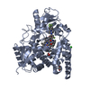 4c27C  4c28C  4c0cS S: Starting model for refinement C: citing same article ( |
|---|---|
| Similar structure data |


-Links
 PDBj
PDBj
- Assembly
| Deposited unit | 
| |||||||||
|---|---|---|---|---|---|---|---|---|---|---|
| 1 | 
| |||||||||
| Unit cell |
| |||||||||
| Components on special symmetry positions |
|
-Components
| #1: Protein | / TC14DM / CYTOCHROME P450 51 / LANOSTEROL 14-ALPHA DEMETHYLASE / STEROL 14-DEMETHYLASE / CYP51 Mass: 53286.820 Da / Num. of mol.: 1 / Fragment: UNP RESIDUES 32-481 Source method: isolated from a genetically manipulated source Source: (gene. exp.) TRYPANOSOMA CRUZI (eukaryote) / Plasmid: PCW / Production host:  ESCHERICHIA COLI (E. coli) / Strain (production host): BL21(DE3) / Variant (production host): HMS174 ESCHERICHIA COLI (E. coli) / Strain (production host): BL21(DE3) / Variant (production host): HMS174References: UniProt: Q5I4E1, UniProt: Q7Z1V1*PLUS, sterol 14alpha-demethylase | ||||
|---|---|---|---|---|---|
| #2: Chemical | ChemComp-HEM / Heme B  Mass: 616.487 Da / Num. of mol.: 1 / Source method: obtained synthetically / Formula: C34H32FeN4O4 Mass: 616.487 Da / Num. of mol.: 1 / Source method: obtained synthetically / Formula: C34H32FeN4O4 | ||||
| #3: Chemical | ChemComp-J5Y /   Mass: 611.108 Da / Num. of mol.: 1 / Source method: obtained synthetically / Formula: C34H32ClFN6O2 Mass: 611.108 Da / Num. of mol.: 1 / Source method: obtained synthetically / Formula: C34H32ClFN6O2 | ||||
| #4: Chemical | ChemComp-SO4 / Sulfate  Mass: 96.063 Da / Num. of mol.: 4 / Source method: obtained synthetically / Formula: SO4 Mass: 96.063 Da / Num. of mol.: 4 / Source method: obtained synthetically / Formula: SO4#5: Water | ChemComp-HOH / | Water Mass: 18.015 Da / Num. of mol.: 33 / Source method: isolated from a natural source / Formula: H2O Mass: 18.015 Da / Num. of mol.: 33 / Source method: isolated from a natural source / Formula: H2OSequence details | FIRST 32 RESIDUES AT THE N-TERMINUS ARE REPLACED WITH THE MAKKTSSKGKL SEQUENCE, 6XHIS TAG ...FIRST 32 RESIDUES AT THE N-TERMINUS ARE REPLACED WITH THE MAKKTSSKGK | |
-Experimental details
-Experiment
| Experiment | Method: X-RAY DIFFRACTION / Number of used crystals: 1 |
|---|
- Sample preparation
| Crystal | Density Matthews: 2.58 Å3/Da / Density % sol: 52.42 % / Description: NONE |
|---|---|
| Crystal grow | pH: 5.6 Details: 0.1 M SODIUM ACETATE PH 5.6, 0.25 M AMMONIUM SULFATE, 25% PEG 3350 |
-Data collection
| Diffraction | Mean temperature: 110 K |
|---|---|
| Diffraction source | Source: SYNCHROTRON / Site: ALS  / Beamline: 8.3.1 / Wavelength: 1.11587 / Beamline: 8.3.1 / Wavelength: 1.11587 |
| Detector | Type: MARRESERCH / Detector: CCD / Date: Jul 30, 2014 / Details: MIRRORS |
| Radiation | Monochromator: SI(111) / Protocol: SINGLE WAVELENGTH / Monochromatic (M) / Laue (L): M / Scattering type: x-ray |
| Radiation wavelength | Wavelength: 1.11587 Å / Relative weight: 1 |
| Reflection | Resolution: 2.48→117.78 Å / Num. obs: 20811 / % possible obs: 100 % / Observed criterion σ(I): 0.5 / Redundancy: 15.2 % / Biso Wilson estimate: 55.7 Å2 / Rmerge(I) obs: 0.22 / Net I/σ(I): 11.5 |
| Reflection shell | Resolution: 2.48→2.61 Å / Redundancy: 15.6 % / Mean I/σ(I) obs: 1.8 / % possible all: 100 |
- Processing
| Software |
| ||||||||||||||||||||||||||||||||||||||||||||||||||||||||||||||||||||||||||||||||||||||||||||||||||||||||||||||||||||||||||||||||||||||||||||||||||||||||||||||||||||||||||||||||||||||
|---|---|---|---|---|---|---|---|---|---|---|---|---|---|---|---|---|---|---|---|---|---|---|---|---|---|---|---|---|---|---|---|---|---|---|---|---|---|---|---|---|---|---|---|---|---|---|---|---|---|---|---|---|---|---|---|---|---|---|---|---|---|---|---|---|---|---|---|---|---|---|---|---|---|---|---|---|---|---|---|---|---|---|---|---|---|---|---|---|---|---|---|---|---|---|---|---|---|---|---|---|---|---|---|---|---|---|---|---|---|---|---|---|---|---|---|---|---|---|---|---|---|---|---|---|---|---|---|---|---|---|---|---|---|---|---|---|---|---|---|---|---|---|---|---|---|---|---|---|---|---|---|---|---|---|---|---|---|---|---|---|---|---|---|---|---|---|---|---|---|---|---|---|---|---|---|---|---|---|---|---|---|---|---|
| Refinement | Method to determine structure: MOLECULAR REPLACEMENT Starting model: PDB ENTRY 4C0C Resolution: 2.48→110.95 Å / Cor.coef. Fo:Fc: 0.951 / Cor.coef. Fo:Fc free: 0.926 / SU B: 9.855 / SU ML: 0.212 / Cross valid method: THROUGHOUT / ESU R: 0.383 / ESU R Free: 0.269 / Stereochemistry target values: MAXIMUM LIKELIHOOD / Details: HYDROGENS HAVE BEEN ADDED IN THE RIDING POSITIONS.
| ||||||||||||||||||||||||||||||||||||||||||||||||||||||||||||||||||||||||||||||||||||||||||||||||||||||||||||||||||||||||||||||||||||||||||||||||||||||||||||||||||||||||||||||||||||||
| Solvent computation | Ion probe radii: 0.8 Å / Shrinkage radii: 0.8 Å / VDW probe radii: 1.2 Å / Solvent model: MASK | ||||||||||||||||||||||||||||||||||||||||||||||||||||||||||||||||||||||||||||||||||||||||||||||||||||||||||||||||||||||||||||||||||||||||||||||||||||||||||||||||||||||||||||||||||||||
| Displacement parameters | Biso mean: 41.053 Å2
| ||||||||||||||||||||||||||||||||||||||||||||||||||||||||||||||||||||||||||||||||||||||||||||||||||||||||||||||||||||||||||||||||||||||||||||||||||||||||||||||||||||||||||||||||||||||
| Refinement step | Cycle: LAST / Resolution: 2.48→110.95 Å
| ||||||||||||||||||||||||||||||||||||||||||||||||||||||||||||||||||||||||||||||||||||||||||||||||||||||||||||||||||||||||||||||||||||||||||||||||||||||||||||||||||||||||||||||||||||||
| Refine LS restraints |
|