 Movie
Movie Controller


+ Open data

- Basic information
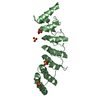
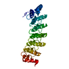
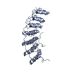
| Entry | Database: PDB / ID: 4uuc | ||||||
|---|---|---|---|---|---|---|---|
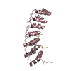
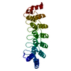
| Title | Crystal structure of human ASB11 ankyrin repeat domain | ||||||
 Components Components | ANKYRIN REPEAT AND SOCS BOX PROTEIN 11 | ||||||
 Keywords Keywords |  SIGNALING PROTEIN SIGNALING PROTEIN | ||||||
| Function / homology |  Function and homology informationpost-translational protein modification / positive regulation of protein catabolic process / Antigen processing: Ubiquitination & Proteasome degradation / Neddylation / protein ubiquitination / intracellular signal transduction / endoplasmic reticulum / cytosol Function and homology informationpost-translational protein modification / positive regulation of protein catabolic process / Antigen processing: Ubiquitination & Proteasome degradation / Neddylation / protein ubiquitination / intracellular signal transduction / endoplasmic reticulum / cytosolSimilarity search - Function | ||||||
| Biological species |  HOMO SAPIENS (human) HOMO SAPIENS (human) | ||||||
| Method | X-RAY DIFFRACTION / SYNCHROTRON / MOLECULAR REPLACEMENT / Resolution: 1.8 Å | ||||||
 Authors Authors | Pinkas, D.M. / Sanvitale, C. / Kragh Nielsen, T. / Guo, K. / Sorrell, F. / Berridge, G. / Ayinampudi, V. / Wang, D. / Newman, J.A. / Tallant, C. ...Pinkas, D.M. / Sanvitale, C. / Kragh Nielsen, T. / Guo, K. / Sorrell, F. / Berridge, G. / Ayinampudi, V. / Wang, D. / Newman, J.A. / Tallant, C. / Chaikuad, A. / Canning, P. / Kopec, J. / Krojer, T. / Vollmar, M. / Allerston, C.K. / Chalk, R. / Burgess-Brown, N. / von Delft, F. / Arrowsmith, C.H. / Edwards, A. / Bountra, C. / Bullock, A. | ||||||
 Citation Citation | Journal: To be Published Title: Crystal Structure of Human Asb11 Ankyrin Repeat Domain Authors: Pinkas, D.M. / Sanvitale, C. / Kragh Nielsen, T. / Guo, K. / Sorrell, F. / Berridge, G. / Ayinampudi, V. / Wang, D. / Newman, J.A. / Tallant, C. / Chaikuad, A. / Canning, P. / Kopec, J. / ...Authors: Pinkas, D.M. / Sanvitale, C. / Kragh Nielsen, T. / Guo, K. / Sorrell, F. / Berridge, G. / Ayinampudi, V. / Wang, D. / Newman, J.A. / Tallant, C. / Chaikuad, A. / Canning, P. / Kopec, J. / Krojer, T. / Vollmar, M. / Allerston, C.K. / Chalk, R. / Burgess-Brown, N. / von Delft, F. / Arrowsmith, C.H. / Edwards, A. / Bountra, C. / Bullock, A. | ||||||
| History |
|
- Structure visualization
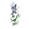
| Structure viewer | Molecule: MolmilJmol/JSmol |
|---|
- Downloads & links
-Download
| PDBx/mmCIF format | 4uuc.cif.gz | 92.9 KB | Display | PDBx/mmCIF format |
|---|---|---|---|---|
| PDB format | pdb4uuc.ent.gz | 71.8 KB | Display | PDB format |
| PDBx/mmJSON format | 4uuc.json.gz | Tree view | PDBx/mmJSON format | |
| Others |  Other downloads Other downloads |
-Validation report
| Arichive directory | https://data.pdbj.org/pub/pdb/validation_reports/uu/4uucftp://data.pdbj.org/pub/pdb/validation_reports/uu/4uuc | HTTPS FTP |
|---|
-Related structure data
| Related structure data | 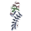 3zkjS S: Starting model for refinement |
|---|---|
| Similar structure data |


-Links
 PDBj
PDBj- Assembly
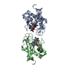
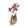
| Deposited unit | 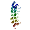
| ||||||||
|---|---|---|---|---|---|---|---|---|---|
| 1 |
| ||||||||
| Unit cell |
|
-Components
| #1: Protein | Mass: 23998.590 Da / Num. of mol.: 1 / Fragment: ANKYRIN REPEAT DOMAIN, RESIDUES 64-287 Source method: isolated from a genetically manipulated source Source: (gene. exp.) HOMO SAPIENS (human) / Production host:  ESCHERICHIA COLI (E. coli) / Strain (production host): BL21 / References: UniProt: Q8WXH4 ESCHERICHIA COLI (E. coli) / Strain (production host): BL21 / References: UniProt: Q8WXH4 |
|---|---|
| #2: Water | ChemComp-HOH / Water Mass: 18.015 Da / Num. of mol.: 243 / Source method: isolated from a natural source / Formula: H2O Mass: 18.015 Da / Num. of mol.: 243 / Source method: isolated from a natural source / Formula: H2O |
-Experimental details
-Experiment
| Experiment | Method: X-RAY DIFFRACTION / Number of used crystals: 1 |
|---|
- Sample preparation
| Crystal | Density Matthews: 2.6 Å3/Da / Density % sol: 52.72 % / Description: NONE |
|---|---|
| Crystal grow | pH: 5.5 Details: 0.2M AMMONIUM ACETATE, 25% PEG3350, 0.1M BIS-TRIS PH 5.5 |
-Data collection
| Diffraction | Mean temperature: 100 K |
|---|---|
| Diffraction source | Source: SYNCHROTRON / Site: Diamond  / Beamline: I03 / Wavelength: 0.97625 / Beamline: I03 / Wavelength: 0.97625 |
| Detector | Type: DECTRIS PILATUS 6M / Detector: PIXEL / Date: Jul 6, 2014 |
| Radiation | Protocol: SINGLE WAVELENGTH / Monochromatic (M) / Laue (L): M / Scattering type: x-ray |
| Radiation wavelength | Wavelength: 0.97625 Å / Relative weight: 1 |
| Reflection | Resolution: 1.8→89.6 Å / Num. obs: 23795 / % possible obs: 99.7 % / Observed criterion σ(I): -3 / Redundancy: 5.9 % / Biso Wilson estimate: 22.8 Å2 / Rmerge(I) obs: 0.1 / Net I/σ(I): 11.1 |
| Reflection shell | Resolution: 1.8→1.84 Å / Redundancy: 3 % / Rmerge(I) obs: 0.73 / Mean I/σ(I) obs: 2.3 / % possible all: 98.7 |
- Processing
| Software |
| |||||||||||||||||||||||||||||||||||||||||||||||||||||||||||||||
|---|---|---|---|---|---|---|---|---|---|---|---|---|---|---|---|---|---|---|---|---|---|---|---|---|---|---|---|---|---|---|---|---|---|---|---|---|---|---|---|---|---|---|---|---|---|---|---|---|---|---|---|---|---|---|---|---|---|---|---|---|---|---|---|---|
| Refinement | Method to determine structure: MOLECULAR REPLACEMENT Starting model: PDB ENTRY 3ZKJ Resolution: 1.8→52.281 Å / SU ML: 0.19 / σ(F): 1.35 / Phase error: 18.89 / Stereochemistry target values: ML
| |||||||||||||||||||||||||||||||||||||||||||||||||||||||||||||||
| Solvent computation | Shrinkage radii: 0.9 Å / VDW probe radii: 1.11 Å / Solvent model: FLAT BULK SOLVENT MODEL | |||||||||||||||||||||||||||||||||||||||||||||||||||||||||||||||
| Displacement parameters | Biso mean: 26.86 Å2 | |||||||||||||||||||||||||||||||||||||||||||||||||||||||||||||||
| Refinement step | Cycle: LAST / Resolution: 1.8→52.281 Å
| |||||||||||||||||||||||||||||||||||||||||||||||||||||||||||||||
| Refine LS restraints |
| |||||||||||||||||||||||||||||||||||||||||||||||||||||||||||||||
| LS refinement shell |
|
