 Movie
Movie Controller

[English] 日本語
 Yorodumi
- PDB-4uu9: Crystal structure of the human c5a in complex with MEDI7814 a neu... -
+ Open data

- Basic information
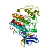
| Entry | Database: PDB / ID: 4uu9 | ||||||
|---|---|---|---|---|---|---|---|
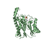
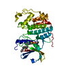
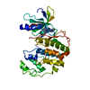
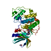
| Title | Crystal structure of the human c5a in complex with MEDI7814 a neutralising antibody | ||||||
 Components Components |
| ||||||
 Keywords Keywords |  IMMUNE SYSTEM / FV / COMPLEMENT SYSTEM IMMUNE SYSTEM / FV / COMPLEMENT SYSTEM | ||||||
| Function / homology |  Function and homology information Function and homology informationTerminal pathway of complement / membrane attack complex / Activation of C3 and C5 / negative regulation of macrophage chemotaxis / complement activation, alternative pathway / chemokine activity / endopeptidase inhibitor activity / positive regulation of vascular endothelial growth factor production / positive regulation of chemokine production / complement activation, classical pathway ...Terminal pathway of complement / membrane attack complex / Activation of C3 and C5 / negative regulation of macrophage chemotaxis / complement activation, alternative pathway / chemokine activity / endopeptidase inhibitor activity / positive regulation of vascular endothelial growth factor production / positive regulation of chemokine production / complement activation, classical pathway / Peptide ligand-binding receptors / Regulation of Complement cascade / chemotaxis / G alpha (i) signalling events / killing of cells of another organism / cell surface receptor signaling pathway / inflammatory response / G protein-coupled receptor signaling pathway / signaling receptor binding / extracellular space / extracellular exosome / extracellular regionSimilarity search - Function | ||||||
| Biological species |  HOMO SAPIENS (human) HOMO SAPIENS (human) | ||||||
| Method | X-RAY DIFFRACTION / SYNCHROTRON / MOLECULAR REPLACEMENT / Resolution: 2.12 Å | ||||||
 Authors Authors | Colley, C. / Sridharan, S. / Dobson, C. / Popovic, B. / Debreczeni, J.E. / Hargreaves, D. / Edwards, B. / Brennan, J. / England, L. / Fung, S. ...Colley, C. / Sridharan, S. / Dobson, C. / Popovic, B. / Debreczeni, J.E. / Hargreaves, D. / Edwards, B. / Brennan, J. / England, L. / Fung, S. / An Eghobamien, L. / Sivars, U. / Woods, R. / Flavell, L. / Renshaw, G.J. / Wickson, K. / Wilkinson, T. / Davies, R. / Bonnell, J. / Warrener, P. / Howes, R. / Vaughan, T. | ||||||
 Citation Citation | Journal: MAbs / Year: 2018 Title: Structure and characterization of a high affinity C5a monoclonal antibody that blocks binding to C5aR1 and C5aR2 receptors. Authors: Colley, C.S. / Popovic, B. / Sridharan, S. / Debreczeni, J.E. / Hargeaves, D. / Fung, M. / An, L.L. / Edwards, B. / Arnold, J. / England, E. / Eghobamien, L. / Sivars, U. / Flavell, L. / ...Authors: Colley, C.S. / Popovic, B. / Sridharan, S. / Debreczeni, J.E. / Hargeaves, D. / Fung, M. / An, L.L. / Edwards, B. / Arnold, J. / England, E. / Eghobamien, L. / Sivars, U. / Flavell, L. / Renshaw, J. / Wickson, K. / Warrener, P. / Zha, J. / Bonnell, J. / Woods, R. / Wilkinson, T. / Dobson, C. / Vaughan, T.J. | ||||||
| History |
|
- Structure visualization
| Structure viewer | Molecule: MolmilJmol/JSmol |
|---|
- Downloads & links
-Download
| PDBx/mmCIF format | 4uu9.cif.gz | 233.4 KB | Display | PDBx/mmCIF format |
|---|---|---|---|---|
| PDB format | pdb4uu9.ent.gz | 197.9 KB | Display | PDB format |
| PDBx/mmJSON format | 4uu9.json.gz | Tree view | PDBx/mmJSON format | |
| Others |  Other downloads Other downloads |
-Validation report
| Arichive directory | https://data.pdbj.org/pub/pdb/validation_reports/uu/4uu9ftp://data.pdbj.org/pub/pdb/validation_reports/uu/4uu9 | HTTPS FTP |
|---|
-Related structure data
| Similar structure data |
|---|


-Links
 PDBj
PDBj


- Assembly
| Deposited unit | 
| ||||||||
|---|---|---|---|---|---|---|---|---|---|
| 1 | 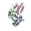
| ||||||||
| 2 | 
| ||||||||
| Unit cell |
|
-Components
| #1: Antibody | Mass: 13508.873 Da / Num. of mol.: 2 / Fragment: VARIABLE DOMAIN HEAVY CHAIN Source method: isolated from a genetically manipulated source Source: (gene. exp.) HOMO SAPIENS (human) / Description: HUMANISED ANTIBODY FRAGMENT / Production host:  ESCHERICHIA COLI BL21 (bacteria) ESCHERICHIA COLI BL21 (bacteria)#2: Antibody | Mass: 11629.880 Da / Num. of mol.: 2 / Fragment: VARIABLE DOMAIN LIGHT CHAIN Source method: isolated from a genetically manipulated source Source: (gene. exp.) HOMO SAPIENS (human) / Description: HUMANISED ANTIBODY FRAGMENT / Production host: ESCHERICHIA COLI BL21 (bacteria)#3: Protein | Complement component 5 / C5A ANAPHYLATOXIN C5A / C3 AND PZP-LIKE ALPHA-2-MACROGLOBULIN DOMAIN-CONTAINING PROTEIN 4Mass: 8375.754 Da / Num. of mol.: 2 / Fragment: C5A Source method: isolated from a genetically manipulated source Source: (gene. exp.) HOMO SAPIENS (human) / Description: HUMANISED ANTIBODY FRAGMENT / Production host: ESCHERICHIA COLI BL21 (bacteria) / References: UniProt: P01031#4: Chemical | ChemComp-SO4 / Sulfate  Mass: 96.063 Da / Num. of mol.: 4 / Source method: obtained synthetically / Formula: SO4 Mass: 96.063 Da / Num. of mol.: 4 / Source method: obtained synthetically / Formula: SO4#5: Water | ChemComp-HOH / | Water Mass: 18.015 Da / Num. of mol.: 301 / Source method: isolated from a natural source / Formula: H2O Mass: 18.015 Da / Num. of mol.: 301 / Source method: isolated from a natural source / Formula: H2O |
|---|
-Experimental details
-Experiment
| Experiment | Method: X-RAY DIFFRACTION / Number of used crystals: 1 |
|---|
- Sample preparation
| Crystal | Density Matthews: 2.41 Å3/Da / Density % sol: 49 % / Description: NONE |
|---|---|
| Crystal grow | Details: 20% PEG10K, 8% ETHYLENE GLYCOL, 100MM HEPES PH 7.5 |
-Data collection
| Diffraction | Mean temperature: 100 K |
|---|---|
| Diffraction source | Source: SYNCHROTRON / Site: ESRF  / Beamline: ID23-1 / Wavelength: 0.976 / Beamline: ID23-1 / Wavelength: 0.976 |
| Detector | Type: ADSC CCD / Detector: CCD / Date: Nov 13, 2010 |
| Radiation | Protocol: SINGLE WAVELENGTH / Monochromatic (M) / Laue (L): M / Scattering type: x-ray |
| Radiation wavelength | Wavelength: 0.976 Å / Relative weight: 1 |
| Reflection | Resolution: 2.12→126 Å / Num. obs: 48925 / % possible obs: 100 % / Observed criterion σ(I): 2 / Redundancy: 14.2 % / Rmerge(I) obs: 0.13 / Net I/σ(I): 19 |
| Reflection shell | Resolution: 2.12→2.24 Å / Redundancy: 14.7 % / Rmerge(I) obs: 0.59 / Mean I/σ(I) obs: 8.4 / % possible all: 100 |
- Processing
| Software |
| ||||||||||||||||||||||||||||||||||||||||||||||||||||||||||||||||||||||||||||||||||||||||||||||||||||||||||||||||||||||||||||||||||||||||||||||||||||||||||||||||||||||||||||||||||||||
|---|---|---|---|---|---|---|---|---|---|---|---|---|---|---|---|---|---|---|---|---|---|---|---|---|---|---|---|---|---|---|---|---|---|---|---|---|---|---|---|---|---|---|---|---|---|---|---|---|---|---|---|---|---|---|---|---|---|---|---|---|---|---|---|---|---|---|---|---|---|---|---|---|---|---|---|---|---|---|---|---|---|---|---|---|---|---|---|---|---|---|---|---|---|---|---|---|---|---|---|---|---|---|---|---|---|---|---|---|---|---|---|---|---|---|---|---|---|---|---|---|---|---|---|---|---|---|---|---|---|---|---|---|---|---|---|---|---|---|---|---|---|---|---|---|---|---|---|---|---|---|---|---|---|---|---|---|---|---|---|---|---|---|---|---|---|---|---|---|---|---|---|---|---|---|---|---|---|---|---|---|---|---|---|
| Refinement | Method to determine structure: MOLECULAR REPLACEMENT / Resolution: 2.12→86.07 Å / Cor.coef. Fo:Fc: 0.95 / Cor.coef. Fo:Fc free: 0.929 / SU B: 6.54 / SU ML: 0.09 / Cross valid method: THROUGHOUT / ESU R: 0.158 / ESU R Free: 0.14 / Stereochemistry target values: MAXIMUM LIKELIHOOD Details: HYDROGENS HAVE BEEN ADDED IN THE RIDING POSITIONS. U VALUES WITH TLS ADDED
| ||||||||||||||||||||||||||||||||||||||||||||||||||||||||||||||||||||||||||||||||||||||||||||||||||||||||||||||||||||||||||||||||||||||||||||||||||||||||||||||||||||||||||||||||||||||
| Solvent computation | Ion probe radii: 0.8 Å / Shrinkage radii: 0.8 Å / VDW probe radii: 1.2 Å / Solvent model: MASK | ||||||||||||||||||||||||||||||||||||||||||||||||||||||||||||||||||||||||||||||||||||||||||||||||||||||||||||||||||||||||||||||||||||||||||||||||||||||||||||||||||||||||||||||||||||||
| Displacement parameters | Biso mean: 28.421 Å2
| ||||||||||||||||||||||||||||||||||||||||||||||||||||||||||||||||||||||||||||||||||||||||||||||||||||||||||||||||||||||||||||||||||||||||||||||||||||||||||||||||||||||||||||||||||||||
| Refinement step | Cycle: LAST / Resolution: 2.12→86.07 Å
| ||||||||||||||||||||||||||||||||||||||||||||||||||||||||||||||||||||||||||||||||||||||||||||||||||||||||||||||||||||||||||||||||||||||||||||||||||||||||||||||||||||||||||||||||||||||
| Refine LS restraints |
|