 Movie
Movie Controller


+ Open data

- Basic information
| Entry | Database: PDB / ID: 4r7b | ||||||
|---|---|---|---|---|---|---|---|
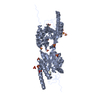
| Title | Crystal structure of pneumococcal LicA in complex with choline | ||||||
 Components Components | Choline kinase | ||||||
 Keywords Keywords | TRANSFERASE / protein kinase-like fold | ||||||
| Function / homology |  Function and homology informationcholine kinase activity / kinase activity / phosphorylation / nucleotide binding Function and homology informationcholine kinase activity / kinase activity / phosphorylation / nucleotide bindingSimilarity search - Function | ||||||
| Biological species |   Streptococcus pneumoniae (bacteria) Streptococcus pneumoniae (bacteria) | ||||||
| Method | X-RAY DIFFRACTION / SYNCHROTRON / MOLECULAR REPLACEMENT / Resolution: 2.01 Å | ||||||
 Authors Authors | Wang, L. / Jiang, Y.L. / Zhou, C.Z. / Chen, Y.X. | ||||||
 Citation Citation | Journal: Plos One / Year: 2015 Title: Structural and enzymatic characterization of the choline kinase LicA from Streptococcus pneumoniae Authors: Wang, L. / Jiang, Y.L. / Zhang, J.R. / Zhou, C.Z. / Chen, Y.X. | ||||||
| History |
|
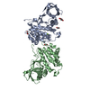
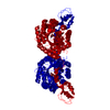
- Structure visualization
| Structure viewer | Molecule: MolmilJmol/JSmol |
|---|
- Downloads & links
-Download
| PDBx/mmCIF format | 4r7b.cif.gz | 133.9 KB | Display | PDBx/mmCIF format |
|---|---|---|---|---|
| PDB format | pdb4r7b.ent.gz | 103.4 KB | Display | PDB format |
| PDBx/mmJSON format | 4r7b.json.gz | Tree view | PDBx/mmJSON format | |
| Others |  Other downloads Other downloads |
-Validation report
| Arichive directory | https://data.pdbj.org/pub/pdb/validation_reports/r7/4r7bftp://data.pdbj.org/pub/pdb/validation_reports/r7/4r7b | HTTPS FTP |
|---|
-Related structure data
| Related structure data | 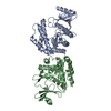 4r77SC 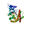 4r78C S: Starting model for refinement C: citing same article ( |
|---|---|
| Similar structure data |


-Links
 PDBj
PDBj- Assembly
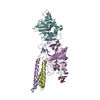
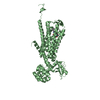
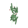
| Deposited unit | 
| ||||||||
|---|---|---|---|---|---|---|---|---|---|
| 1 |
| ||||||||
| 2 | 
| ||||||||
| 3 | 
| ||||||||
| Unit cell |
|
-Components
| #1: Protein | Mass: 35610.867 Da / Num. of mol.: 2 Source method: isolated from a genetically manipulated source Source: (gene. exp.) Streptococcus pneumoniae (bacteria) / Gene: licA / Production host: Escherichia coli (E. coli)References: UniProt: Q93MI3, UniProt: Q8DPI4*PLUS, choline kinase#2: Chemical | Choline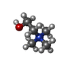  Mass: 104.171 Da / Num. of mol.: 2 / Source method: obtained synthetically / Formula: C5H14NO Mass: 104.171 Da / Num. of mol.: 2 / Source method: obtained synthetically / Formula: C5H14NO#3: Water | ChemComp-HOH / | Water Mass: 18.015 Da / Num. of mol.: 312 / Source method: isolated from a natural source / Formula: H2O Mass: 18.015 Da / Num. of mol.: 312 / Source method: isolated from a natural source / Formula: H2O |
|---|
-Experimental details
-Experiment
| Experiment | Method: X-RAY DIFFRACTION / Number of used crystals: 1 |
|---|
- Sample preparation
| Crystal | Density Matthews: 2.36 Å3/Da / Density % sol: 47.78 % |
|---|---|
| Crystal grow | Temperature: 289 K / Method: vapor diffusion, hanging drop / pH: 7.5 Details: 0.1M HEPES, 1.2M sodium citrate, 4% glycerol, pH 7.5, VAPOR DIFFUSION, HANGING DROP, temperature 289K |
-Data collection
| Diffraction | Mean temperature: 100 K |
|---|---|
| Diffraction source | Source: SYNCHROTRON / Site: SSRF  / Beamline: BL17U / Wavelength: 0.97924 Å / Beamline: BL17U / Wavelength: 0.97924 Å |
| Detector | Type: ADSC QUANTUM 315r / Detector: CCD / Date: Dec 15, 2013 |
| Radiation | Monochromator: Si(111) / Protocol: SINGLE WAVELENGTH / Monochromatic (M) / Laue (L): M / Scattering type: x-ray |
| Radiation wavelength | Wavelength: 0.97924 Å / Relative weight: 1 |
| Reflection | Resolution: 2.01→50 Å / Num. all: 44656 / Num. obs: 44656 / % possible obs: 98.2 % / Observed criterion σ(F): 0 / Observed criterion σ(I): -3 |
| Reflection shell | Resolution: 2.01→2.08 Å / Redundancy: 3.4 % / Rmerge(I) obs: 0.571 / Mean I/σ(I) obs: 3.362 / Rsym value: 0.571 / % possible all: 98 |
- Processing
| Software |
| |||||||||||||||||||||||||||||||||||||||||||||||||||||||||||||||||
|---|---|---|---|---|---|---|---|---|---|---|---|---|---|---|---|---|---|---|---|---|---|---|---|---|---|---|---|---|---|---|---|---|---|---|---|---|---|---|---|---|---|---|---|---|---|---|---|---|---|---|---|---|---|---|---|---|---|---|---|---|---|---|---|---|---|---|
| Refinement | Method to determine structure: MOLECULAR REPLACEMENT Starting model: 4R77 Resolution: 2.01→29.78 Å / Cor.coef. Fo:Fc: 0.954 / Cor.coef. Fo:Fc free: 0.936 / SU B: 4.171 / SU ML: 0.116 / Cross valid method: THROUGHOUT / ESU R: 0.196 / ESU R Free: 0.168 / Stereochemistry target values: MAXIMUM LIKELIHOOD / Details: HYDROGENS HAVE BEEN ADDED IN THE RIDING POSITIONS
| |||||||||||||||||||||||||||||||||||||||||||||||||||||||||||||||||
| Solvent computation | Ion probe radii: 0.8 Å / Shrinkage radii: 0.8 Å / VDW probe radii: 1.4 Å / Solvent model: MASK | |||||||||||||||||||||||||||||||||||||||||||||||||||||||||||||||||
| Displacement parameters | Biso mean: 36.236 Å2
| |||||||||||||||||||||||||||||||||||||||||||||||||||||||||||||||||
| Refinement step | Cycle: LAST / Resolution: 2.01→29.78 Å
| |||||||||||||||||||||||||||||||||||||||||||||||||||||||||||||||||
| Refine LS restraints |
| |||||||||||||||||||||||||||||||||||||||||||||||||||||||||||||||||
| LS refinement shell | Resolution: 2.011→2.063 Å / Total num. of bins used: 20
|
