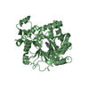
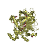
 Movie
Movie Controller


+ Open data

- Basic information
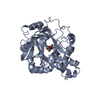
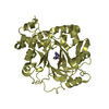
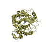
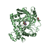
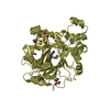
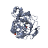
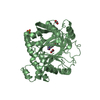
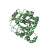
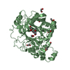
| Entry | Database: PDB / ID: 4ura | ||||||
|---|---|---|---|---|---|---|---|
| Title | Crystal structure of human JMJD2A in complex with compound 14a | ||||||
 Components Components | LYSINE-SPECIFIC DEMETHYLASE 4A | ||||||
 Keywords Keywords |  OXIDOREDUCTASE / JUMONJIC / HISTONE DEMETHYLASE OXIDOREDUCTASE / JUMONJIC / HISTONE DEMETHYLASE | ||||||
| Function / homology |  Function and homology information Function and homology information[histone H3]-trimethyl-L-lysine36 demethylase / histone H3K36me2/H3K36me3 demethylase activity / apoptotic chromosome condensation / histone H3K36 demethylase activity / cardiac muscle hypertrophy in response to stress / histone H3K9me2/H3K9me3 demethylase activity / [histone H3]-trimethyl-L-lysine9 demethylase / histone H3K9 demethylase activity / negative regulation of astrocyte differentiation / histone demethylase activity ...[histone H3]-trimethyl-L-lysine36 demethylase / histone H3K36me2/H3K36me3 demethylase activity / apoptotic chromosome condensation / histone H3K36 demethylase activity / cardiac muscle hypertrophy in response to stress / histone H3K9me2/H3K9me3 demethylase activity / [histone H3]-trimethyl-L-lysine9 demethylase / histone H3K9 demethylase activity / negative regulation of astrocyte differentiation / histone demethylase activity / pericentric heterochromatin / NR1H3 & NR1H2 regulate gene expression linked to cholesterol transport and efflux / methylated histone binding / positive regulation of neuron differentiation / response to nutrient levels / negative regulation of autophagy / HDMs demethylate histones / fibrillar center / Recruitment and ATM-mediated phosphorylation of repair and signaling proteins at DNA double strand breaks / regulation of gene expression / chromatin remodeling / negative regulation of gene expression / negative regulation of DNA-templated transcription / ubiquitin protein ligase binding / chromatin / positive regulation of gene expression / zinc ion binding / nucleoplasm / nucleus / cytosolSimilarity search - Function | ||||||
| Biological species |  HOMO SAPIENS (human) HOMO SAPIENS (human) | ||||||
| Method | X-RAY DIFFRACTION / SYNCHROTRON / OTHER / Resolution: 2.23 Å | ||||||
 Authors Authors | Krojer, T. / England, K.S. / Vollmar, M. / Crawley, L. / Williams, E. / Riesebos, E. / Szykowska, A. / Burgess-Brown, N. / Oppermann, U. / Brennan, P.E. ...Krojer, T. / England, K.S. / Vollmar, M. / Crawley, L. / Williams, E. / Riesebos, E. / Szykowska, A. / Burgess-Brown, N. / Oppermann, U. / Brennan, P.E. / Bountra, C. / Arrowsmith, C.H. / Edwards, A. / von Delft, F. | ||||||
 Citation Citation | Journal: Medchemcomm / Year: 2014 Title: Optimisation of a triazolopyridine based histone demethylase inhibitor yields a potent and selective KDM2A (FBXL11) inhibitor. Authors: England, K.S. / Tumber, A. / Krojer, T. / Scozzafava, G. / Ng, S.S. / Daniel, M. / Szykowska, A. / Che, K. / von Delft, F. / Burgess-Brown, N.A. / Kawamura, A. / Schofield, C.J. / Brennan, P.E. | ||||||
| History |
|
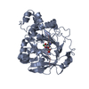
- Structure visualization
| Structure viewer | Molecule: MolmilJmol/JSmol |
|---|
- Downloads & links
-Download
| PDBx/mmCIF format | 4ura.cif.gz | 154.5 KB | Display | PDBx/mmCIF format |
|---|---|---|---|---|
| PDB format | pdb4ura.ent.gz | 121.2 KB | Display | PDB format |
| PDBx/mmJSON format | 4ura.json.gz | Tree view | PDBx/mmJSON format | |
| Others |  Other downloads Other downloads |
-Validation report
| Arichive directory | https://data.pdbj.org/pub/pdb/validation_reports/ur/4uraftp://data.pdbj.org/pub/pdb/validation_reports/ur/4ura | HTTPS FTP |
|---|
-Related structure data
| Similar structure data |
|---|

-Links
 PDBj
PDBj


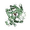
- Assembly
| Deposited unit | 
| ||||||||
|---|---|---|---|---|---|---|---|---|---|
| 1 | 
| ||||||||
| 2 | 
| ||||||||
| Unit cell |
|
-Components
-Protein , 1 types, 2 molecules AB
| #1: Protein | Mass: 41854.617 Da / Num. of mol.: 2 / Fragment: RESIDUES 1-359 Source method: isolated from a genetically manipulated source Source: (gene. exp.) HOMO SAPIENS (human) / Production host:  ESCHERICHIA COLI (E. coli) ESCHERICHIA COLI (E. coli)References: UniProt: O75164, Oxidoreductases; Acting on paired donors, with incorporation or reduction of molecular oxygen; With 2-oxoglutarate as one donor, and incorporation of one atom of oxygen into each donor |
|---|
-Non-polymers , 6 types, 194 molecules 


| #2: Chemical | ChemComp-EDO / Ethylene glycol Mass: 62.068 Da / Num. of mol.: 1 / Source method: obtained synthetically / Formula: C2H6O2 Mass: 62.068 Da / Num. of mol.: 1 / Source method: obtained synthetically / Formula: C2H6O2 | ||||||||
|---|---|---|---|---|---|---|---|---|---|
| #3: Chemical | Sulfate Mass: 96.063 Da / Num. of mol.: 2 / Source method: obtained synthetically / Formula: SO4 Mass: 96.063 Da / Num. of mol.: 2 / Source method: obtained synthetically / Formula: SO4#4: Chemical | Nickel Mass: 58.693 Da / Num. of mol.: 2 / Source method: obtained synthetically / Formula: Ni Mass: 58.693 Da / Num. of mol.: 2 / Source method: obtained synthetically / Formula: Ni#5: Chemical |  Mass: 65.409 Da / Num. of mol.: 2 / Source method: obtained synthetically / Formula: Zn Mass: 65.409 Da / Num. of mol.: 2 / Source method: obtained synthetically / Formula: Zn#6: Chemical |  Mass: 190.159 Da / Num. of mol.: 2 / Source method: obtained synthetically / Formula: C8H6N4O2 Mass: 190.159 Da / Num. of mol.: 2 / Source method: obtained synthetically / Formula: C8H6N4O2#7: Water | ChemComp-HOH / | WaterMass: 18.015 Da / Num. of mol.: 185 / Source method: isolated from a natural source / Formula: H2O |
-Experimental details
-Experiment
| Experiment | Method: X-RAY DIFFRACTION / Number of used crystals: 1 |
|---|
- Sample preparation
| Crystal | Density Matthews: 2.71 Å3/Da / Density % sol: 54.24 % / Description: NONE |
|---|---|
| Crystal grow | pH: 5.9 Details: 0.1M BIS-TRIS PH 5.9, 0.15M AMMONIUM SULFATE, 11% PEG3350 |
-Data collection
| Diffraction | Mean temperature: 100 K |
|---|---|
| Diffraction source | Source: SYNCHROTRON / Site: Diamond  / Beamline: I04-1 / Wavelength: 0.92 / Beamline: I04-1 / Wavelength: 0.92 |
| Detector | Type: DECTRIS PILATUS 6M / Detector: PIXEL / Date: Aug 7, 2014 |
| Radiation | Protocol: SINGLE WAVELENGTH / Monochromatic (M) / Laue (L): M / Scattering type: x-ray |
| Radiation wavelength | Wavelength: 0.92 Å / Relative weight: 1 |
| Reflection | Resolution: 2.23→60.13 Å / Num. obs: 43239 / % possible obs: 99.2 % / Observed criterion σ(I): -3 / Redundancy: 6.7 % / Biso Wilson estimate: 39.62 Å2 / Rmerge(I) obs: 0.09 / Net I/σ(I): 15.9 |
| Reflection shell | Resolution: 2.23→2.29 Å / Redundancy: 6.3 % / Rmerge(I) obs: 0.83 / Mean I/σ(I) obs: 2.3 / % possible all: 97.6 |
- Processing
| Software |
| ||||||||||||||||||||||||||||||||||||||||||||||||||||||||||||||||||||||||||||||||||||||||||||||||||
|---|---|---|---|---|---|---|---|---|---|---|---|---|---|---|---|---|---|---|---|---|---|---|---|---|---|---|---|---|---|---|---|---|---|---|---|---|---|---|---|---|---|---|---|---|---|---|---|---|---|---|---|---|---|---|---|---|---|---|---|---|---|---|---|---|---|---|---|---|---|---|---|---|---|---|---|---|---|---|---|---|---|---|---|---|---|---|---|---|---|---|---|---|---|---|---|---|---|---|---|
| Refinement | Method to determine structure: OTHER Starting model: NONE Resolution: 2.23→60.134 Å / SU ML: 0.33 / σ(F): 1.36 / Phase error: 28.91 / Stereochemistry target values: ML
| ||||||||||||||||||||||||||||||||||||||||||||||||||||||||||||||||||||||||||||||||||||||||||||||||||
| Solvent computation | Shrinkage radii: 0.9 Å / VDW probe radii: 1.11 Å / Solvent model: FLAT BULK SOLVENT MODEL | ||||||||||||||||||||||||||||||||||||||||||||||||||||||||||||||||||||||||||||||||||||||||||||||||||
| Refinement step | Cycle: LAST / Resolution: 2.23→60.134 Å
| ||||||||||||||||||||||||||||||||||||||||||||||||||||||||||||||||||||||||||||||||||||||||||||||||||
| Refine LS restraints |
| ||||||||||||||||||||||||||||||||||||||||||||||||||||||||||||||||||||||||||||||||||||||||||||||||||
| LS refinement shell |
|
