 Movie
Movie Controller

[English] 日本語
 Yorodumi
- PDB-4um1: Engineered Ls-AChBP with alpha4-alpha4 binding pocket in complex ... -
+ Open data

- Basic information
| Entry | Database: PDB / ID: 4um1 | ||||||
|---|---|---|---|---|---|---|---|
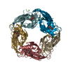
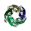
| Title | Engineered Ls-AChBP with alpha4-alpha4 binding pocket in complex with NS3573 | ||||||
 Components Components | ACETYLCHOLINE-BINDING PROTEIN | ||||||
 Keywords Keywords |  SIGNALING PROTEIN / ION CHANNEL / RECEPTOR STOICHIOMETRY / CYS-LOOP RECEPTOR / ACETYLCHOLINE BINDING PROTEIN SIGNALING PROTEIN / ION CHANNEL / RECEPTOR STOICHIOMETRY / CYS-LOOP RECEPTOR / ACETYLCHOLINE BINDING PROTEIN | ||||||
| Function / homology |  Function and homology informationacetylcholine receptor activity / acetylcholine-gated monoatomic cation-selective channel activity / synaptic cleft / response to nicotine / neuron projection / synapse / membrane Function and homology informationacetylcholine receptor activity / acetylcholine-gated monoatomic cation-selective channel activity / synaptic cleft / response to nicotine / neuron projection / synapse / membraneSimilarity search - Function | ||||||
| Biological species |   LYMNAEA STAGNALIS (great pond snail) LYMNAEA STAGNALIS (great pond snail) | ||||||
| Method | X-RAY DIFFRACTION / SYNCHROTRON / MOLECULAR REPLACEMENT / Resolution: 2.83 Å | ||||||
 Authors Authors | Shahsavar, A. / Kastrup, J.S. / Balle, T. / Gajhede, M. | ||||||
 Citation Citation | Journal: Mol.Pharmacol. / Year: 2015 Title: Achbp Engineered to Mimic the Alpha4-Alpha4 Binding Pocket in Alpha4Beta2 Nicotinic Acetylcholine Receptors Reveals Interface Specific Interactions Important for Binding and Activity Authors: Shahsavar, A. / Ahring, P.K. / Olsen, J.A. / Krintel, C. / Kastrup, J.S. / Balle, T. / Gajhede, M. | ||||||
| History |
|
- Structure visualization
| Structure viewer | Molecule: MolmilJmol/JSmol |
|---|
- Downloads & links
-Download
| PDBx/mmCIF format | 4um1.cif.gz | 211 KB | Display | PDBx/mmCIF format |
|---|---|---|---|---|
| PDB format | pdb4um1.ent.gz | 171.1 KB | Display | PDB format |
| PDBx/mmJSON format | 4um1.json.gz | Tree view | PDBx/mmJSON format | |
| Others |  Other downloads Other downloads |
-Validation report
| Arichive directory | https://data.pdbj.org/pub/pdb/validation_reports/um/4um1ftp://data.pdbj.org/pub/pdb/validation_reports/um/4um1 | HTTPS FTP |
|---|
-Related structure data
| Related structure data |  4um3C  3u8kS C: citing same article ( S: Starting model for refinement |
|---|---|
| Similar structure data |


-Links
 PDBj
PDBj

- Assembly
| Deposited unit | 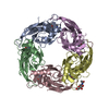
| ||||||||
|---|---|---|---|---|---|---|---|---|---|
| 1 |
| ||||||||
| Unit cell |
|
-Components
| #1: Protein | Mass: 26053.143 Da / Num. of mol.: 5 / Mutation: YES Source method: isolated from a genetically manipulated source Details: 1-(5-ETHOXYPYRIDIN-3-YL)-1,4-DIAZEPANE (NS3573) BINDS AT THE INTERFACE OF EACH TWO MONOMERS Source: (gene. exp.) LYMNAEA STAGNALIS (great pond snail) / Plasmid: PFASTBAC / Production host:   SPODOPTERA FRUGIPERDA (fall armyworm) / Strain (production host): SF9 / References: UniProt: P58154 SPODOPTERA FRUGIPERDA (fall armyworm) / Strain (production host): SF9 / References: UniProt: P58154#2: Chemical | ChemComp-09P /   Mass: 221.299 Da / Num. of mol.: 5 / Source method: obtained synthetically / Formula: C12H19N3O Mass: 221.299 Da / Num. of mol.: 5 / Source method: obtained synthetically / Formula: C12H19N3O#3: Sugar | ChemComp-NAG / | N-Acetylglucosamine  Type: D-saccharide, beta linking / Mass: 221.208 Da / Num. of mol.: 1 Type: D-saccharide, beta linking / Mass: 221.208 Da / Num. of mol.: 1Source method: isolated from a genetically manipulated source Formula: C8H15NO6 #4: Water | ChemComp-HOH / | Water Mass: 18.015 Da / Num. of mol.: 116 / Source method: isolated from a natural source / Formula: H2O Mass: 18.015 Da / Num. of mol.: 116 / Source method: isolated from a natural source / Formula: H2O |
|---|
-Experimental details
-Experiment
| Experiment | Method: X-RAY DIFFRACTION / Number of used crystals: 1 |
|---|
- Sample preparation
| Crystal | Density Matthews: 2.43 Å3/Da / Density % sol: 49.34 % / Description: NONE |
|---|---|
| Crystal grow | pH: 8 Details: 0.1 M TRIS BASE (PH 8.0), 1-3% V/V POLYETHYLENE GLYCOL (PEG) 400 AND 1.8-2.3 M (NH4)2SO4 |
-Data collection
| Diffraction | Mean temperature: 100 K |
|---|---|
| Diffraction source | Source: SYNCHROTRON / Site: ESRF  / Beamline: ID23-2 / Wavelength: 1 / Beamline: ID23-2 / Wavelength: 1 |
| Detector | Type: MARMOSAIC 225 mm CCD / Detector: CCD / Details: MIRRORS |
| Radiation | Monochromator: SI(111) / Protocol: SINGLE WAVELENGTH / Monochromatic (M) / Laue (L): M / Scattering type: x-ray |
| Radiation wavelength | Wavelength: 1 Å / Relative weight: 1 |
| Reflection | Resolution: 2.83→49.18 Å / Num. obs: 29761 / % possible obs: 100 % / Observed criterion σ(I): 0 / Redundancy: 7.8 % / Biso Wilson estimate: 51.25 Å2 / Rmerge(I) obs: 0.21 / Net I/σ(I): 10.5 |
| Reflection shell | Resolution: 2.83→2.98 Å / Redundancy: 7.9 % / Rmerge(I) obs: 1.37 / Mean I/σ(I) obs: 2 / % possible all: 100 |
- Processing
| Software |
| |||||||||||||||||||||||||||||||||||||||||||||||||||||||||||||||||||||||||||||||||||||||||||||||||||||||||||||||||||||||||||||||||||||||||||||||||||
|---|---|---|---|---|---|---|---|---|---|---|---|---|---|---|---|---|---|---|---|---|---|---|---|---|---|---|---|---|---|---|---|---|---|---|---|---|---|---|---|---|---|---|---|---|---|---|---|---|---|---|---|---|---|---|---|---|---|---|---|---|---|---|---|---|---|---|---|---|---|---|---|---|---|---|---|---|---|---|---|---|---|---|---|---|---|---|---|---|---|---|---|---|---|---|---|---|---|---|---|---|---|---|---|---|---|---|---|---|---|---|---|---|---|---|---|---|---|---|---|---|---|---|---|---|---|---|---|---|---|---|---|---|---|---|---|---|---|---|---|---|---|---|---|---|---|---|---|---|
| Refinement | Method to determine structure: MOLECULAR REPLACEMENT Starting model: PDB ENTRY 3U8K Resolution: 2.83→49.178 Å / SU ML: 0.39 / σ(F): 0.16 / Phase error: 23.85 / Stereochemistry target values: ML
| |||||||||||||||||||||||||||||||||||||||||||||||||||||||||||||||||||||||||||||||||||||||||||||||||||||||||||||||||||||||||||||||||||||||||||||||||||
| Solvent computation | Shrinkage radii: 0.9 Å / VDW probe radii: 1.11 Å / Solvent model: FLAT BULK SOLVENT MODEL | |||||||||||||||||||||||||||||||||||||||||||||||||||||||||||||||||||||||||||||||||||||||||||||||||||||||||||||||||||||||||||||||||||||||||||||||||||
| Displacement parameters | Biso mean: 35 Å2 | |||||||||||||||||||||||||||||||||||||||||||||||||||||||||||||||||||||||||||||||||||||||||||||||||||||||||||||||||||||||||||||||||||||||||||||||||||
| Refinement step | Cycle: LAST / Resolution: 2.83→49.178 Å
| |||||||||||||||||||||||||||||||||||||||||||||||||||||||||||||||||||||||||||||||||||||||||||||||||||||||||||||||||||||||||||||||||||||||||||||||||||
| Refine LS restraints |
| |||||||||||||||||||||||||||||||||||||||||||||||||||||||||||||||||||||||||||||||||||||||||||||||||||||||||||||||||||||||||||||||||||||||||||||||||||
| LS refinement shell |
|
