Mass: 18.015 Da / Num. of mol.: 547 / Source method: isolated from a natural source / Formula: H2O
-
Experimental details
-
Experiment
Experiment
Method: X-RAY DIFFRACTION / Number of used crystals: 1
-
Sample preparation
Crystal
Density Matthews: 3.51 Å3/Da / Density % sol: 64.96 %
Crystal grow
Temperature: 282 K / Method: vapor diffusion, sitting drop / pH: 7.8 Details: 3 M ammonium sulfate, 10% (v/v) glycerol, 75 mM sodium chloride, 12.5 mM tris (hydroxymethyl) amino methane, 1.25 mM ethylenediaminetetraacetic acid.
Resolution: 1.67→34.3 Å / Cor.coef. Fo:Fc: 0.971 / Cor.coef. Fo:Fc free: 0.962 / SU B: 4.115 / SU ML: 0.058 / Cross valid method: THROUGHOUT / ESU R: 0.087 / ESU R Free: 0.072 / Stereochemistry target values: MAXIMUM LIKELIHOOD / Details: HYDROGENS HAVE BEEN ADDED IN THE RIDING POSITIONS
Rfactor
Num. reflection
% reflection
Selection details
Rfree
0.18047
4330
4.9 %
RANDOM
Rwork
0.1562
-
-
-
obs
0.15736
84703
99.92 %
-
Solvent computation
Ion probe radii: 0.8 Å / Shrinkage radii: 0.8 Å / VDW probe radii: 1.4 Å / Solvent model: MASK
Displacement parameters
Biso mean: 30.121 Å2
Baniso -1
Baniso -2
Baniso -3
1-
0.87 Å2
0.43 Å2
-0 Å2
2-
-
0.87 Å2
0 Å2
3-
-
-
-2.82 Å2
Refinement step
Cycle: 1 / Resolution: 1.67→34.3 Å
Protein
Nucleic acid
Ligand
Solvent
Total
Num. atoms
3799
0
164
547
4510
Refine LS restraints
Refine-ID
Type
Dev ideal
Dev ideal target
Number
X-RAY DIFFRACTION
r_bond_refined_d
0.008
0.019
4231
X-RAY DIFFRACTION
r_angle_refined_deg
1.229
1.987
5800
LS refinement shell
Resolution: 1.67→1.713 Å / Total num. of bins used: 20
Rfactor
Num. reflection
% reflection
Rfree
0.273
297
-
Rwork
0.243
6172
-
obs
-
-
99.95 %
Refinement TLS params.
Method: refined / Refine-ID: X-RAY DIFFRACTION
ID
L11 (°2)
L12 (°2)
L13 (°2)
L22 (°2)
L23 (°2)
L33 (°2)
S11 (Å °)
S12 (Å °)
S13 (Å °)
S21 (Å °)
S22 (Å °)
S23 (Å °)
S31 (Å °)
S32 (Å °)
S33 (Å °)
T11 (Å2)
T12 (Å2)
T13 (Å2)
T22 (Å2)
T23 (Å2)
T33 (Å2)
Origin x (Å)
Origin y (Å)
Origin z (Å)
1
0.0533
0.0175
0.0187
0.0937
-0.0035
0.01
-0.0015
-0.0015
0.0009
-0.0116
-0.0013
0.0073
-0.0039
-0.0032
0.0028
0.0124
0.0035
-0.0009
0.0059
-0.0013
0.064
35.3708
-17.0159
-37.586
2
29.3776
12.2532
22.7864
9.6981
11.8034
22.5275
0.9704
-1.5761
-0.0163
0.9612
-0.8174
0.18
1.3274
-1.55
-0.153
0.1412
-0.083
0.0066
0.1121
0.017
0.041
15.4178
-20.3485
-36.6257
3
0.613
0.6335
-0.1677
2.832
-0.239
0.0479
-0.005
0.0067
-0.0051
0.0488
0.0057
0.0247
-0.0015
-0.002
-0.0007
0.0205
0.0058
-0.0009
0.0069
-0.0015
0.0583
37.8533
-14.6007
-32.0403
4
0.1571
-0.0293
0.0205
0.2042
0.2097
0.2439
-0.0193
-0.0776
-0.0985
0.0242
-0.0052
0.0726
0.0511
-0.0245
0.0245
0.0952
0.0333
-0.0432
0.0542
0.0217
0.1229
42.0903
-12.1645
-41.0314
5
0.0856
-0.0034
-0.0212
0.1643
-0.0637
0.1416
-0.0019
-0.0185
0.002
-0.0156
0.0008
0.006
0.0099
-0.0091
0.0011
0.0197
0.0068
-0.0043
0.0087
-0.004
0.0739
37.6745
-17.388
-38.6462
6
4.866
-3.0404
3.6427
8.7364
7.9558
18.0404
0.1116
0.1464
-0.086
-0.1503
-0.2682
0.1968
-0.0414
-0.1573
0.1567
0.09
0.0043
-0.0029
0.0873
0.0333
0.1419
18.2901
-8.99
-44.6567
Refinement TLS group
ID
Refine-ID
Refine TLS-ID
Auth asym-ID
Auth seq-ID
1
X-RAY DIFFRACTION
1
A
-3 - 478
2
X-RAY DIFFRACTION
2
B
1001
3
X-RAY DIFFRACTION
3
C
1002
4
X-RAY DIFFRACTION
4
D
1001 - 1014
5
X-RAY DIFFRACTION
5
H
1 - 547
6
X-RAY DIFFRACTION
6
E
1
+
About Yorodumi
-
News
-
Feb 9, 2022. New format data for meta-information of EMDB entries
New format data for meta-information of EMDB entries
Version 3 of the EMDB header file is now the official format.
The previous official version 1.9 will be removed from the archive.
In the structure databanks used in Yorodumi, some data are registered as the other names, "COVID-19 virus" and "2019-nCoV". Here are the details of the virus and the list of structure data.
Jan 31, 2019. EMDB accession codes are about to change! (news from PDBe EMDB page)
EMDB accession codes are about to change! (news from PDBe EMDB page)
The allocation of 4 digits for EMDB accession codes will soon come to an end. Whilst these codes will remain in use, new EMDB accession codes will include an additional digit and will expand incrementally as the available range of codes is exhausted. The current 4-digit format prefixed with “EMD-” (i.e. EMD-XXXX) will advance to a 5-digit format (i.e. EMD-XXXXX), and so on. It is currently estimated that the 4-digit codes will be depleted around Spring 2019, at which point the 5-digit format will come into force.
The EM Navigator/Yorodumi systems omit the EMD- prefix.
Related info.:Q: What is EMD? / ID/Accession-code notation in Yorodumi/EM Navigator
Yorodumi is a browser for structure data from EMDB, PDB, SASBDB, etc.
This page is also the successor to EM Navigator detail page, and also detail information page/front-end page for Omokage search.
The word "yorodu" (or yorozu) is an old Japanese word meaning "ten thousand". "mi" (miru) is to see.
Related info.:EMDB / PDB / SASBDB / Comparison of 3 databanks / Yorodumi Search / Aug 31, 2016. New EM Navigator & Yorodumi / Yorodumi Papers / Jmol/JSmol / Function and homology information / Changes in new EM Navigator and Yorodumi
 Movie
Movie Controller

 Yorodumi
 Open data
 Basic information
 Components
 Keywords
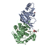
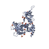
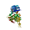
 LYASE /
  Function and homology information
 Authors
 Citation
 Structure visualization
 Downloads & links
 Other downloads
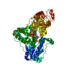


 PDBj
PDBj


 Assembly
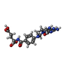


 Mass: 457.440 Da / Num. of mol.: 1 / Source method: obtained synthetically / Formula: C20H23N7O6
 Mass: 785.550 Da / Num. of mol.: 1 / Source method: obtained synthetically / Formula: C27H33N9O15P2 / Comment: FAD*YM
 Mass: 96.063 Da / Num. of mol.: 14 / Source method: obtained synthetically / Formula: SO4
 Mass: 122.143 Da / Num. of mol.: 1 / Source method: obtained synthetically / Formula: C4H12NO3 / Comment: pH buffer*YM
 Sample preparation
 / Beamline: 14.1 / Wavelength: 0.91842 Å
 Processing