 Movie
Movie Controller

[English] 日本語
 Yorodumi
- PDB-4u0m: Structure of the Vibrio cholerae di-nucleotide cyclase (DncV) mut... -
+ Open data

- Basic information
| Entry | Database: PDB / ID: 4u0m | |||||||||
|---|---|---|---|---|---|---|---|---|---|---|
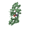
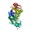
| Title | Structure of the Vibrio cholerae di-nucleotide cyclase (DncV) mutant D193N in complex with ATP, GTP and 5MTHFGLU2 | |||||||||
 Components Components | Cyclic AMP-GMP synthase | |||||||||
 Keywords Keywords |  TRANSFERASE / regulation / mutation TRANSFERASE / regulation / mutation | |||||||||
| Function / homology |  Function and homology information Function and homology information3',3'-cyclic GMP-AMP synthase activity / diguanylate cyclase activity / cyclic nucleotide biosynthetic process / negative regulation of chemotaxis / Transferases; Transferring phosphorus-containing groups; Nucleotidyltransferases / defense response to virus / GTP binding / ATP binding / metal ion bindingSimilarity search - Function | |||||||||
| Biological species |  Vibrio cholerae El Tor N16961 (bacteria) Vibrio cholerae El Tor N16961 (bacteria) | |||||||||
| Method | X-RAY DIFFRACTION / SYNCHROTRON / MOLECULAR REPLACEMENT / Resolution: 2.3 Å | |||||||||
 Authors Authors | Zhu, D. / Xiang, Y. | |||||||||
| Funding support |  China, 2items China, 2items
| |||||||||
 Citation Citation | Journal: Mol.Cell / Year: 2014 Title: Structural Biochemistry of a Vibrio cholerae Dinucleotide Cyclase Reveals Cyclase Activity Regulation by Folates. Authors: Zhu, D. / Wang, L. / Shang, G. / Liu, X. / Zhu, J. / Lu, D. / Wang, L. / Kan, B. / Zhang, J.R. / Xiang, Y. | |||||||||
| History |
|
- Structure visualization
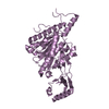
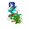
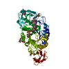
| Structure viewer | Molecule: MolmilJmol/JSmol |
|---|
- Downloads & links
-Download
| PDBx/mmCIF format | 4u0m.cif.gz | 350.8 KB | Display | PDBx/mmCIF format |
|---|---|---|---|---|
| PDB format | pdb4u0m.ent.gz | 284.4 KB | Display | PDB format |
| PDBx/mmJSON format | 4u0m.json.gz | Tree view | PDBx/mmJSON format | |
| Others |  Other downloads Other downloads |
-Validation report
| Arichive directory | https://data.pdbj.org/pub/pdb/validation_reports/u0/4u0mftp://data.pdbj.org/pub/pdb/validation_reports/u0/4u0m | HTTPS FTP |
|---|
-Related structure data
| Related structure data |  4u03C  4u0lSC  4u0nC C: citing same article ( S: Starting model for refinement |
|---|---|
| Similar structure data |


-Links
 PDBj
PDBj- Assembly
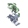
| Deposited unit | 
| ||||||||
|---|---|---|---|---|---|---|---|---|---|
| 1 | 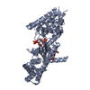
| ||||||||
| 2 | 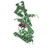
| ||||||||
| Unit cell |
|
-Components
-Protein , 1 types, 2 molecules AB
| #1: Protein | Mass: 48824.410 Da / Num. of mol.: 2 / Fragment: UNP residues 1-419 / Mutation: D193N Source method: isolated from a genetically manipulated source Source: (gene. exp.) Vibrio cholerae El Tor N16961 (bacteria)Gene: dncV, VC_0179 / Plasmid: pET22b / Production host: Escherichia coli (E. coli) / References: UniProt: Q9KVG7, cyclic GMP-AMP synthase |
|---|
-Non-polymers , 5 types, 384 molecules 


| #2: Chemical | Guanosine triphosphate Mass: 523.180 Da / Num. of mol.: 2 / Source method: obtained synthetically / Formula: C10H16N5O14P3 / Comment: GTP, energy-carrying molecule*YM Mass: 523.180 Da / Num. of mol.: 2 / Source method: obtained synthetically / Formula: C10H16N5O14P3 / Comment: GTP, energy-carrying molecule*YM#3: Chemical | Adenosine triphosphate Mass: 507.181 Da / Num. of mol.: 2 / Source method: obtained synthetically / Formula: C10H16N5O13P3 / Comment: ATP, energy-carrying molecule*YM Mass: 507.181 Da / Num. of mol.: 2 / Source method: obtained synthetically / Formula: C10H16N5O13P3 / Comment: ATP, energy-carrying molecule*YM#4: Chemical |  Mass: 24.305 Da / Num. of mol.: 2 / Source method: obtained synthetically / Formula: Mg Mass: 24.305 Da / Num. of mol.: 2 / Source method: obtained synthetically / Formula: Mg#5: Chemical | ChemComp-TLL / |  Mass: 588.570 Da / Num. of mol.: 1 / Source method: obtained synthetically / Formula: C25H32N8O9 Mass: 588.570 Da / Num. of mol.: 1 / Source method: obtained synthetically / Formula: C25H32N8O9#6: Water | ChemComp-HOH / | WaterMass: 18.015 Da / Num. of mol.: 377 / Source method: isolated from a natural source / Formula: H2O |
|---|
-Experimental details
-Experiment
| Experiment | Method: X-RAY DIFFRACTION / Number of used crystals: 1 |
|---|
- Sample preparation
| Crystal | Density Matthews: 2.22 Å3/Da / Density % sol: 44.63 % |
|---|---|
| Crystal grow | Temperature: 293 K / Method: vapor diffusion, hanging drop / pH: 7 Details: 20% w/v PEG 3350, 200mM NaCl, 100mM Tris-HCl, 5mM GTP, 5mM ATP |
-Data collection
| Diffraction | Mean temperature: 100 K |
|---|---|
| Diffraction source | Source: SYNCHROTRON / Site: SSRF / Beamline: BL17U / Wavelength: 0.968 Å |
| Detector | Type: ADSC QUANTUM 315r / Detector: CCD / Date: May 20, 2013 |
| Radiation | Protocol: SINGLE WAVELENGTH / Monochromatic (M) / Laue (L): M / Scattering type: x-ray |
| Radiation wavelength | Wavelength: 0.968 Å / Relative weight: 1 |
| Reflection | Resolution: 2.3→103.7 Å / Num. obs: 38293 / % possible obs: 99.5 % / Redundancy: 3.3 % / Net I/σ(I): 12.9 |
- Processing
| Software | Name: PHENIX / Version: (phenix.refine: 1.8.2_1309) / Classification: refinement | |||||||||||||||||||||||||||||||||||||||||||||||||||||||||||||||||||||||||||||||||||||||||||||||||||||||||
|---|---|---|---|---|---|---|---|---|---|---|---|---|---|---|---|---|---|---|---|---|---|---|---|---|---|---|---|---|---|---|---|---|---|---|---|---|---|---|---|---|---|---|---|---|---|---|---|---|---|---|---|---|---|---|---|---|---|---|---|---|---|---|---|---|---|---|---|---|---|---|---|---|---|---|---|---|---|---|---|---|---|---|---|---|---|---|---|---|---|---|---|---|---|---|---|---|---|---|---|---|---|---|---|---|---|---|
| Refinement | Method to determine structure: MOLECULAR REPLACEMENT Starting model: 4U0L Resolution: 2.3→42.568 Å / SU ML: 0.32 / Cross valid method: FREE R-VALUE / σ(F): 1.34 / Phase error: 27.34 / Stereochemistry target values: ML
| |||||||||||||||||||||||||||||||||||||||||||||||||||||||||||||||||||||||||||||||||||||||||||||||||||||||||
| Solvent computation | Shrinkage radii: 0.9 Å / VDW probe radii: 1.11 Å / Solvent model: FLAT BULK SOLVENT MODEL | |||||||||||||||||||||||||||||||||||||||||||||||||||||||||||||||||||||||||||||||||||||||||||||||||||||||||
| Refinement step | Cycle: LAST / Resolution: 2.3→42.568 Å
| |||||||||||||||||||||||||||||||||||||||||||||||||||||||||||||||||||||||||||||||||||||||||||||||||||||||||
| Refine LS restraints |
| |||||||||||||||||||||||||||||||||||||||||||||||||||||||||||||||||||||||||||||||||||||||||||||||||||||||||
| LS refinement shell |
| |||||||||||||||||||||||||||||||||||||||||||||||||||||||||||||||||||||||||||||||||||||||||||||||||||||||||
| Refinement TLS params. | Method: refined / Origin x: 65.6854 Å / Origin y: 8.3343 Å / Origin z: 177.149 Å
| |||||||||||||||||||||||||||||||||||||||||||||||||||||||||||||||||||||||||||||||||||||||||||||||||||||||||
| Refinement TLS group | Selection details: all |
