 Movie
Movie Controller


+ Open data

- Basic information
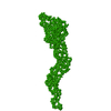
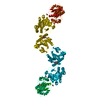
| Entry | Database: PDB / ID: 4tt1 | ||||||
|---|---|---|---|---|---|---|---|
| Title | Crystal structure of fragment 1600-1733 of HSV1 UL36, native | ||||||
 Components Components | Deneddylase | ||||||
 Keywords Keywords |  HYDROLASE / Fibrous protein / Tegument Protein / VIRAL PROTEIN / PROTEIN FIBRIL HYDROLASE / Fibrous protein / Tegument Protein / VIRAL PROTEIN / PROTEIN FIBRIL | ||||||
| Function / homology |  Function and homology information Function and homology informationnuclear capsid assembly / deNEDDylase activity / viral tegument / symbiont-mediated suppression of cytoplasmic pattern recognition receptor signaling pathway / deubiquitinase activity / viral DNA genome replication / symbiont-mediated perturbation of host ubiquitin-like protein modification / ubiquitinyl hydrolase 1 / cysteine-type deubiquitinase activity / host cell cytoplasm ...nuclear capsid assembly / deNEDDylase activity / viral tegument / symbiont-mediated suppression of cytoplasmic pattern recognition receptor signaling pathway / deubiquitinase activity / viral DNA genome replication / symbiont-mediated perturbation of host ubiquitin-like protein modification / ubiquitinyl hydrolase 1 / cysteine-type deubiquitinase activity / host cell cytoplasm / Hydrolases; Acting on peptide bonds (peptidases); Cysteine endopeptidases / host cell nucleus / proteolysisSimilarity search - Function | ||||||
| Biological species |  Herpes simplex virus Herpes simplex virus | ||||||
| Method | X-RAY DIFFRACTION / SYNCHROTRON / MOLECULAR REPLACEMENT / Resolution: 2.75 Å | ||||||
 Authors Authors | Scrima, N. / Bressanelli, S. / Roche, S. | ||||||
 Citation Citation | Journal: J.Biol.Chem. / Year: 2015 Title: Insights into Herpesvirus Tegument Organization from Structural Analyses of the 970 Central Residues of HSV-1 UL36 Protein. Authors: Scrima, N. / Lepault, J. / Boulard, Y. / Pasdeloup, D. / Bressanelli, S. / Roche, S. | ||||||
| History |
|
- Structure visualization
| Structure viewer | Molecule: MolmilJmol/JSmol |
|---|
- Downloads & links
-Download
| PDBx/mmCIF format | 4tt1.cif.gz | 111.5 KB | Display | PDBx/mmCIF format |
|---|---|---|---|---|
| PDB format | pdb4tt1.ent.gz | 87.8 KB | Display | PDB format |
| PDBx/mmJSON format | 4tt1.json.gz | Tree view | PDBx/mmJSON format | |
| Others |  Other downloads Other downloads |
-Validation report
| Arichive directory | https://data.pdbj.org/pub/pdb/validation_reports/tt/4tt1ftp://data.pdbj.org/pub/pdb/validation_reports/tt/4tt1 | HTTPS FTP |
|---|
-Related structure data
| Related structure data |  4tt0SC S: Starting model for refinement C: citing same article ( |
|---|---|
| Similar structure data |


-Links
 PDBj
PDBj- Assembly
| Deposited unit | 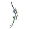
| ||||||||
|---|---|---|---|---|---|---|---|---|---|
| 1 | 
| ||||||||
| Unit cell |
|
-Components
| #1: Protein | Mass: 15093.057 Da / Num. of mol.: 2 / Fragment: Residues 1625-1757 Source method: isolated from a genetically manipulated source Source: (gene. exp.) Herpes simplex virus (type 1 / strain 17)Gene: UL36 / Production host:  Escherichia coli (E. coli) Escherichia coli (E. coli)References: UniProt: P10220, ubiquitinyl hydrolase 1, Hydrolases; Acting on peptide bonds (peptidases); Cysteine endopeptidases#2: Chemical | Phosphate  Mass: 94.971 Da / Num. of mol.: 2 / Source method: obtained synthetically / Formula: PO4 Mass: 94.971 Da / Num. of mol.: 2 / Source method: obtained synthetically / Formula: PO4#3: Water | ChemComp-HOH / | Water Mass: 18.015 Da / Num. of mol.: 9 / Source method: isolated from a natural source / Formula: H2O Mass: 18.015 Da / Num. of mol.: 9 / Source method: isolated from a natural source / Formula: H2O |
|---|
-Experimental details
-Experiment
| Experiment | Method: X-RAY DIFFRACTION |
|---|
- Sample preparation
| Crystal | Density Matthews: 4.65 Å3/Da / Density % sol: 73.52 % |
|---|---|
| Crystal grow | Temperature: 293 K / Method: vapor diffusion, hanging drop / Details: 200 mM diammonium hydrogenophosphate, 20% PEG 3350 |
-Data collection
| Diffraction | Mean temperature: 100 K |
|---|---|
| Diffraction source | Source: SYNCHROTRON / Site: SOLEIL  / Beamline: PROXIMA 1 / Wavelength: 0.97911 Å / Beamline: PROXIMA 1 / Wavelength: 0.97911 Å |
| Detector | Type: PSI PILATUS 6M / Detector: PIXEL / Date: Oct 14, 2012 |
| Radiation | Protocol: SINGLE WAVELENGTH / Monochromatic (M) / Laue (L): M / Scattering type: x-ray |
| Radiation wavelength | Wavelength: 0.97911 Å / Relative weight: 1 |
| Reflection | Resolution: 2.75→38.317 Å / Num. obs: 15531 / % possible obs: 99.9 % / Redundancy: 12.8 % / Net I/σ(I): 21 |
| Reflection shell | Resolution: 2.6→2.75 Å / Redundancy: 12.4 % / Mean I/σ(I) obs: 1.82 |
- Processing
| Software |
| ||||||||||||||||||||||||||||||||||||||||||||||||||||||||||||||||||||||||||||||||||||||||||||||||||||
|---|---|---|---|---|---|---|---|---|---|---|---|---|---|---|---|---|---|---|---|---|---|---|---|---|---|---|---|---|---|---|---|---|---|---|---|---|---|---|---|---|---|---|---|---|---|---|---|---|---|---|---|---|---|---|---|---|---|---|---|---|---|---|---|---|---|---|---|---|---|---|---|---|---|---|---|---|---|---|---|---|---|---|---|---|---|---|---|---|---|---|---|---|---|---|---|---|---|---|---|---|---|
| Refinement | Method to determine structure: MOLECULAR REPLACEMENT Starting model: 4TT0 Resolution: 2.75→38.317 Å / SU ML: 0.44 / Cross valid method: FREE R-VALUE / σ(F): 1.34 / Phase error: 29.93 / Stereochemistry target values: ML
| ||||||||||||||||||||||||||||||||||||||||||||||||||||||||||||||||||||||||||||||||||||||||||||||||||||
| Solvent computation | Shrinkage radii: 0.9 Å / VDW probe radii: 1.11 Å / Solvent model: FLAT BULK SOLVENT MODEL | ||||||||||||||||||||||||||||||||||||||||||||||||||||||||||||||||||||||||||||||||||||||||||||||||||||
| Refinement step | Cycle: LAST / Resolution: 2.75→38.317 Å
| ||||||||||||||||||||||||||||||||||||||||||||||||||||||||||||||||||||||||||||||||||||||||||||||||||||
| Refine LS restraints |
| ||||||||||||||||||||||||||||||||||||||||||||||||||||||||||||||||||||||||||||||||||||||||||||||||||||
| LS refinement shell |
| ||||||||||||||||||||||||||||||||||||||||||||||||||||||||||||||||||||||||||||||||||||||||||||||||||||
| Refinement TLS params. | Method: refined / Refine-ID: X-RAY DIFFRACTION
| ||||||||||||||||||||||||||||||||||||||||||||||||||||||||||||||||||||||||||||||||||||||||||||||||||||
| Refinement TLS group |
|
