 Movie
Movie Controller

[English] 日本語
 Yorodumi
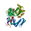
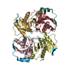
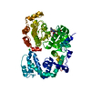
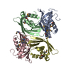
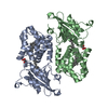
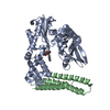
- PDB-4tnb: Crystal Structure of G Protein-Coupled Receptor Kinase 5 in Compl... -
+ Open data

- Basic information
| Entry | Database: PDB / ID: 4tnb | ||||||
|---|---|---|---|---|---|---|---|
| Title | Crystal Structure of G Protein-Coupled Receptor Kinase 5 in Complex with Sangivamycin | ||||||
 Components Components | G protein-coupled receptor kinase 5 | ||||||
 Keywords Keywords |  SIGNALING PROTEIN / GRK5-Sangivamycin complex / GPCR Kinase / Kinase / inhibitor SIGNALING PROTEIN / GRK5-Sangivamycin complex / GPCR Kinase / Kinase / inhibitor | ||||||
| Function / homology |  Function and homology informationG-protein-coupled receptor kinase / beta-adrenergic receptor kinase activity / G protein-coupled receptor kinase activity / tachykinin receptor signaling pathway / regulation of G protein-coupled receptor signaling pathway / fat cell differentiation / regulation of signal transduction / protein kinase C binding / adenylate cyclase-modulating G protein-coupled receptor signaling pathway / phospholipid binding ...G-protein-coupled receptor kinase / beta-adrenergic receptor kinase activity / G protein-coupled receptor kinase activity / tachykinin receptor signaling pathway / regulation of G protein-coupled receptor signaling pathway / fat cell differentiation / regulation of signal transduction / protein kinase C binding / adenylate cyclase-modulating G protein-coupled receptor signaling pathway / phospholipid binding / Wnt signaling pathway / G alpha (s) signalling events / G alpha (q) signalling events / nuclear membrane / protein autophosphorylation / regulation of cell cycle / protein kinase activity / nuclear speck / G protein-coupled receptor signaling pathway / protein serine/threonine kinase activity / apoptotic process / positive regulation of cell population proliferation / negative regulation of apoptotic process / ATP binding / plasma membrane / cytosol / cytoplasm Function and homology informationG-protein-coupled receptor kinase / beta-adrenergic receptor kinase activity / G protein-coupled receptor kinase activity / tachykinin receptor signaling pathway / regulation of G protein-coupled receptor signaling pathway / fat cell differentiation / regulation of signal transduction / protein kinase C binding / adenylate cyclase-modulating G protein-coupled receptor signaling pathway / phospholipid binding ...G-protein-coupled receptor kinase / beta-adrenergic receptor kinase activity / G protein-coupled receptor kinase activity / tachykinin receptor signaling pathway / regulation of G protein-coupled receptor signaling pathway / fat cell differentiation / regulation of signal transduction / protein kinase C binding / adenylate cyclase-modulating G protein-coupled receptor signaling pathway / phospholipid binding / Wnt signaling pathway / G alpha (s) signalling events / G alpha (q) signalling events / nuclear membrane / protein autophosphorylation / regulation of cell cycle / protein kinase activity / nuclear speck / G protein-coupled receptor signaling pathway / protein serine/threonine kinase activity / apoptotic process / positive regulation of cell population proliferation / negative regulation of apoptotic process / ATP binding / plasma membrane / cytosol / cytoplasmSimilarity search - Function | ||||||
| Biological species |  Homo sapiens (human) Homo sapiens (human) | ||||||
| Method | X-RAY DIFFRACTION / SYNCHROTRON / MOLECULAR REPLACEMENT / Resolution: 2.113 Å | ||||||
 Authors Authors | Bhardwaj, A. / Komolov, K.E. / Benovic, J.L. | ||||||
 Citation Citation | Journal: J.Biol.Chem. / Year: 2015 Title: Atomic Structure of G Protein-Coupled Receptor Kinase 5 (GRK5) Reveals Distinct Structural Features Novel for GRKs. Authors: Komolov, K.E. / Bhardwaj, A. / Benovic, J.L. | ||||||
| History |
|
- Structure visualization
| Structure viewer | Molecule: MolmilJmol/JSmol |
|---|
- Downloads & links
-Download
| PDBx/mmCIF format | 4tnb.cif.gz | 237.5 KB | Display | PDBx/mmCIF format |
|---|---|---|---|---|
| PDB format | pdb4tnb.ent.gz | 189.4 KB | Display | PDB format |
| PDBx/mmJSON format | 4tnb.json.gz | Tree view | PDBx/mmJSON format | |
| Others |  Other downloads Other downloads |
-Validation report
| Arichive directory | https://data.pdbj.org/pub/pdb/validation_reports/tn/4tnbftp://data.pdbj.org/pub/pdb/validation_reports/tn/4tnb | HTTPS FTP |
|---|
-Related structure data
| Related structure data |  4tndC  3nynS C: citing same article ( S: Starting model for refinement |
|---|---|
| Similar structure data |


-Links
 PDBj
PDBj


- Assembly
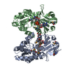
| Deposited unit | 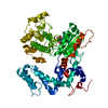
| ||||||||
|---|---|---|---|---|---|---|---|---|---|
| 1 |
| ||||||||
| Unit cell |
|
-Components
| #1: Protein | Mass: 67942.672 Da / Num. of mol.: 1 Source method: isolated from a genetically manipulated source Source: (gene. exp.) Homo sapiens (human) / Gene: GRK5, GPRK5 / Production host:   Spodoptera frugiperda (fall armyworm) Spodoptera frugiperda (fall armyworm)References: UniProt: P34947, G-protein-coupled receptor kinase |
|---|---|
| #2: Chemical | ChemComp-SGV / Sangivamycin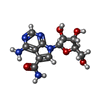  Mass: 309.278 Da / Num. of mol.: 1 / Source method: obtained synthetically / Formula: C12H15N5O5 / Comment: inhibitor*YM Mass: 309.278 Da / Num. of mol.: 1 / Source method: obtained synthetically / Formula: C12H15N5O5 / Comment: inhibitor*YM |
| #3: Water | ChemComp-HOH / Water Mass: 18.015 Da / Num. of mol.: 398 / Source method: isolated from a natural source / Formula: H2O Mass: 18.015 Da / Num. of mol.: 398 / Source method: isolated from a natural source / Formula: H2O |
-Experimental details
-Experiment
| Experiment | Method: X-RAY DIFFRACTION |
|---|
- Sample preparation
| Crystal | Density Matthews: 2.11 Å3/Da / Density % sol: 41.74 % |
|---|---|
| Crystal grow | Temperature: 277 K / Method: vapor diffusion, hanging drop / pH: 5.5 Details: 21% PEG 3350, 0.15 M Sodium Chloride, 0.1 M BIS-TRIS pH 5.5 |
-Data collection
| Diffraction | Mean temperature: 80 K |
|---|---|
| Diffraction source | Source: SYNCHROTRON / Site: NSLS  / Beamline: X6A / Wavelength: 0.9795 Å / Beamline: X6A / Wavelength: 0.9795 Å |
| Detector | Type: ADSC QUANTUM 270 / Detector: CCD / Date: Jun 25, 2013 / Details: Toroidal focusing mirror |
| Radiation | Monochromator: Si(111) channel cut monochromator / Protocol: SINGLE WAVELENGTH / Monochromatic (M) / Laue (L): M / Scattering type: x-ray |
| Radiation wavelength | Wavelength: 0.9795 Å / Relative weight: 1 |
| Reflection | Resolution: 2.113→47.567 Å / Num. obs: 28334 / % possible obs: 83.7 % / Redundancy: 3.6 % / Rsym value: 0.112 / Net I/σ(I): 12.64 |
| Reflection shell | Resolution: 2.2→2.24 Å / Redundancy: 1.6 % / Mean I/σ(I) obs: 1.34 / Rsym value: 0.426 / % possible all: 51.9 |
- Processing
| Software |
| ||||||||||||||||||||||||||||||||||||||||||||||||||||||||||||||||||||||||||||||||||||||||||||||||||||||||||||||||||||||||||||||||||||||||||||||||||||||
|---|---|---|---|---|---|---|---|---|---|---|---|---|---|---|---|---|---|---|---|---|---|---|---|---|---|---|---|---|---|---|---|---|---|---|---|---|---|---|---|---|---|---|---|---|---|---|---|---|---|---|---|---|---|---|---|---|---|---|---|---|---|---|---|---|---|---|---|---|---|---|---|---|---|---|---|---|---|---|---|---|---|---|---|---|---|---|---|---|---|---|---|---|---|---|---|---|---|---|---|---|---|---|---|---|---|---|---|---|---|---|---|---|---|---|---|---|---|---|---|---|---|---|---|---|---|---|---|---|---|---|---|---|---|---|---|---|---|---|---|---|---|---|---|---|---|---|---|---|---|---|---|
| Refinement | Method to determine structure: MOLECULAR REPLACEMENT Starting model: 3NYN Resolution: 2.113→47.567 Å / SU ML: 0.26 / Cross valid method: FREE R-VALUE / σ(F): 1.35 / Phase error: 23.88 / Stereochemistry target values: ML
| ||||||||||||||||||||||||||||||||||||||||||||||||||||||||||||||||||||||||||||||||||||||||||||||||||||||||||||||||||||||||||||||||||||||||||||||||||||||
| Solvent computation | Shrinkage radii: 0.9 Å / VDW probe radii: 1.11 Å / Solvent model: FLAT BULK SOLVENT MODEL | ||||||||||||||||||||||||||||||||||||||||||||||||||||||||||||||||||||||||||||||||||||||||||||||||||||||||||||||||||||||||||||||||||||||||||||||||||||||
| Refinement step | Cycle: LAST / Resolution: 2.113→47.567 Å
| ||||||||||||||||||||||||||||||||||||||||||||||||||||||||||||||||||||||||||||||||||||||||||||||||||||||||||||||||||||||||||||||||||||||||||||||||||||||
| Refine LS restraints |
| ||||||||||||||||||||||||||||||||||||||||||||||||||||||||||||||||||||||||||||||||||||||||||||||||||||||||||||||||||||||||||||||||||||||||||||||||||||||
| LS refinement shell |
| ||||||||||||||||||||||||||||||||||||||||||||||||||||||||||||||||||||||||||||||||||||||||||||||||||||||||||||||||||||||||||||||||||||||||||||||||||||||
| Refinement TLS params. | Method: refined / Refine-ID: X-RAY DIFFRACTION
| ||||||||||||||||||||||||||||||||||||||||||||||||||||||||||||||||||||||||||||||||||||||||||||||||||||||||||||||||||||||||||||||||||||||||||||||||||||||
| Refinement TLS group |
|
