 Movie
Movie Controller

[English] 日本語
 Yorodumi
- PDB-4s0g: Crystal structure of PTPN3 (PTPH1) in complex with Eps15 pTyr849 ... -
+ Open data

- Basic information
| Entry | Database: PDB / ID: 4s0g | ||||||
|---|---|---|---|---|---|---|---|
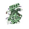
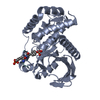
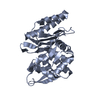
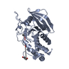
| Title | Crystal structure of PTPN3 (PTPH1) in complex with Eps15 pTyr849 P850V peptide | ||||||
 Components Components |
| ||||||
 Keywords Keywords | HYDROLASE/PROTEIN BINDING / Alpha Beta /  Hydrolase / HYDROLASE-PROTEIN BINDING complex Hydrolase / HYDROLASE-PROTEIN BINDING complex | ||||||
| Function / homology |  Function and homology information Function and homology informationregulation of membrane depolarization during action potential / negative regulation of membrane protein ectodomain proteolysis / Golgi to endosome transport / clathrin coat of coated pit / regulation of sodium ion transmembrane transporter activity / vesicle organization / postsynaptic neurotransmitter receptor internalization / clathrin coat assembly / endocytic recycling / aggresome ...regulation of membrane depolarization during action potential / negative regulation of membrane protein ectodomain proteolysis / Golgi to endosome transport / clathrin coat of coated pit / regulation of sodium ion transmembrane transporter activity / vesicle organization / postsynaptic neurotransmitter receptor internalization / clathrin coat assembly / endocytic recycling / aggresome / endosomal transport / positive regulation of receptor recycling / negative regulation of epidermal growth factor receptor signaling pathway / negative regulation of mitotic cell cycle / sodium channel regulator activity / polyubiquitin modification-dependent protein binding / clathrin-coated pit / cytoskeletal protein binding / phosphotyrosine residue binding / protein dephosphorylation / protein-tyrosine-phosphatase / basal plasma membrane / InlB-mediated entry of Listeria monocytogenes into host cell / protein tyrosine phosphatase activity / liver regeneration / EGFR downregulation / Negative regulation of MET activity / cytoplasmic side of plasma membrane / SH3 domain binding / endocytosis / Negative regulation of MAPK pathway / MAPK cascade / protein transport / Cargo recognition for clathrin-mediated endocytosis / Clathrin-mediated endocytosis / regulation of cell population proliferation / ATPase binding / early endosome membrane / postsynapse / receptor-mediated endocytosis of virus by host cell / cytoskeleton / symbiont entry into host cell / cadherin binding / apical plasma membrane / intracellular membrane-bounded organelle / glutamatergic synapse / calcium ion binding / membrane / plasma membrane / cytosol / cytoplasmSimilarity search - Function | ||||||
| Biological species |  Homo sapiens (human) Homo sapiens (human) | ||||||
| Method | X-RAY DIFFRACTION / SYNCHROTRON / MOLECULAR REPLACEMENT / Resolution: 1.723 Å | ||||||
 Authors Authors | Chen, K.-E. / Meng, T.C. / Wang, A.H.-J. | ||||||
 Citation Citation | Journal: Structure / Year: 2015 Title: Substrate specificity and plasticity of FERM-containing protein tyrosine phosphatases. Authors: Chen, K.E. / Li, M.Y. / Chou, C.C. / Ho, M.R. / Chen, G.C. / Meng, T.C. / Wang, A.H. | ||||||
| History |
|
- Structure visualization
| Structure viewer | Molecule: MolmilJmol/JSmol |
|---|
- Downloads & links
-Download
| PDBx/mmCIF format | 4s0g.cif.gz | 123.3 KB | Display | PDBx/mmCIF format |
|---|---|---|---|---|
| PDB format | pdb4s0g.ent.gz | 95.6 KB | Display | PDB format |
| PDBx/mmJSON format | 4s0g.json.gz | Tree view | PDBx/mmJSON format | |
| Others |  Other downloads Other downloads |
-Validation report
| Arichive directory | https://data.pdbj.org/pub/pdb/validation_reports/s0/4s0gftp://data.pdbj.org/pub/pdb/validation_reports/s0/4s0g | HTTPS FTP |
|---|
-Related structure data
| Related structure data |  4rh5SC  4rh9C  4rhgC  4ri4C  4ri5C S: Starting model for refinement C: citing same article ( |
|---|---|
| Similar structure data |


-Links
 PDBj
PDBj


- Assembly
| Deposited unit | 
| ||||||||
|---|---|---|---|---|---|---|---|---|---|
| 1 |
| ||||||||
| Unit cell |
|
-Components
| #1: Protein | Mass: 34887.664 Da / Num. of mol.: 1 / Fragment: Catalytic domain, UNP residues 628-909 / Mutation: D811A, C842S Source method: isolated from a genetically manipulated source Source: (gene. exp.) Homo sapiens (human) / Gene: PTPH1, PTPN3 / Production host:  Escherichia coli (E. coli) / Strain (production host): BL21 (DE3) / References: UniProt: P26045, protein-tyrosine-phosphatase Escherichia coli (E. coli) / Strain (production host): BL21 (DE3) / References: UniProt: P26045, protein-tyrosine-phosphatase |
|---|---|
| #2: Protein/peptide | / Protein Eps15 / Protein AF-1p Mass: 1126.021 Da / Num. of mol.: 1 / Fragment: Phosphotyrosine 849 peptide, UNP residues 846-854 / Mutation: P850V / Source method: obtained synthetically / Source: (synth.) Homo sapiens (human) / References: UniProt: P42566 |
| #3: Water | ChemComp-HOH / Water Mass: 18.015 Da / Num. of mol.: 137 / Source method: isolated from a natural source / Formula: H2O Mass: 18.015 Da / Num. of mol.: 137 / Source method: isolated from a natural source / Formula: H2O |
-Experimental details
-Experiment
| Experiment | Method: X-RAY DIFFRACTION / Number of used crystals: 1 |
|---|
- Sample preparation
| Crystal | Density Matthews: 1.87 Å3/Da / Density % sol: 34.29 % |
|---|---|
| Crystal grow | Temperature: 293 K / Method: vapor diffusion, sitting drop / pH: 8.5 Details: 0.1M Tris-HCl, 26% PEG 8000, pH 8.5, VAPOR DIFFUSION, SITTING DROP, temperature 293K |
-Data collection
| Diffraction | Mean temperature: 100 K |
|---|---|
| Diffraction source | Source: SYNCHROTRON / Site: SPring-8  / Beamline: BL44XU / Wavelength: 0.9 Å / Beamline: BL44XU / Wavelength: 0.9 Å |
| Detector | Type: RAYONIX MX225HE / Detector: CCD / Date: Jun 2, 2014 |
| Radiation | Monochromator: SAGITALLY FOCUSED Si(111) / Protocol: SINGLE WAVELENGTH / Monochromatic (M) / Laue (L): M / Scattering type: x-ray |
| Radiation wavelength | Wavelength: 0.9 Å / Relative weight: 1 |
| Reflection | Resolution: 1.72→50 Å / Num. all: 28900 / Num. obs: 28900 / % possible obs: 99 % / Observed criterion σ(F): 0 / Observed criterion σ(I): 0 / Redundancy: 3.8 % / Biso Wilson estimate: 19.5 Å2 / Rmerge(I) obs: 0.068 / Net I/σ(I): 15 |
| Reflection shell | Resolution: 1.72→1.78 Å / Redundancy: 3.9 % / Rmerge(I) obs: 0.522 / Mean I/σ(I) obs: 2.1 / Num. unique all: 2839 / % possible all: 99.4 |
- Processing
| Software |
| ||||||||||||||||||||||||||||||||||||||||||||||||||||||||||||||||||
|---|---|---|---|---|---|---|---|---|---|---|---|---|---|---|---|---|---|---|---|---|---|---|---|---|---|---|---|---|---|---|---|---|---|---|---|---|---|---|---|---|---|---|---|---|---|---|---|---|---|---|---|---|---|---|---|---|---|---|---|---|---|---|---|---|---|---|---|
| Refinement | Method to determine structure: MOLECULAR REPLACEMENT Starting model: PDB Entry 4RH5 Resolution: 1.723→37.076 Å / SU ML: 0.21 / σ(F): 1.33 / Phase error: 23.02 / Stereochemistry target values: MLHL
| ||||||||||||||||||||||||||||||||||||||||||||||||||||||||||||||||||
| Solvent computation | Shrinkage radii: 0.9 Å / VDW probe radii: 1.11 Å / Solvent model: FLAT BULK SOLVENT MODEL | ||||||||||||||||||||||||||||||||||||||||||||||||||||||||||||||||||
| Refinement step | Cycle: LAST / Resolution: 1.723→37.076 Å
| ||||||||||||||||||||||||||||||||||||||||||||||||||||||||||||||||||
| Refine LS restraints |
| ||||||||||||||||||||||||||||||||||||||||||||||||||||||||||||||||||
| LS refinement shell | Refine-ID: X-RAY DIFFRACTION / Total num. of bins used: 10
|
