 Movie
Movie Controller

[English] 日本語
 Yorodumi
- PDB-4rzq: Structural Analysis of Substrate, Reaction Intermediate and Produ... -
+ Open data

- Basic information
| Entry | Database: PDB / ID: 4rzq | ||||||
|---|---|---|---|---|---|---|---|
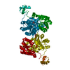
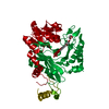
| Title | Structural Analysis of Substrate, Reaction Intermediate and Product Binding in Haemophilus influenzae Biotin Carboxylase | ||||||
 Components Components | Biotin carboxylase | ||||||
 Keywords Keywords | LIGASE / ATP Grasp / Carboxylase / Biotin Carboxyl Carrier Protein and Carboxyltransferase | ||||||
| Function / homology |  Function and homology informationbiotin carboxylase / biotin carboxylase activity / malonyl-CoA biosynthetic process / acetyl-CoA carboxylase activity / fatty acid biosynthetic process / ATP binding / metal ion binding Function and homology informationbiotin carboxylase / biotin carboxylase activity / malonyl-CoA biosynthetic process / acetyl-CoA carboxylase activity / fatty acid biosynthetic process / ATP binding / metal ion bindingSimilarity search - Function | ||||||
| Biological species |  Haemophilus influenzae Rd KW20 (bacteria) Haemophilus influenzae Rd KW20 (bacteria) | ||||||
| Method | X-RAY DIFFRACTION / SYNCHROTRON / MOLECULAR REPLACEMENT / Resolution: 1.98 Å | ||||||
 Authors Authors | Broussard, T.C. / Pakhomova, S. / Neau, D.B. / Champion, T.S. / Bonnot, R. / Waldrop, G.L. | ||||||
 Citation Citation | Journal: Biochemistry / Year: 2015 Title: Structural Analysis of Substrate, Reaction Intermediate, and Product Binding in Haemophilus influenzae Biotin Carboxylase. Authors: Broussard, T.C. / Pakhomova, S. / Neau, D.B. / Bonnot, R. / Waldrop, G.L. | ||||||
| History |
|
- Structure visualization
| Structure viewer | Molecule: MolmilJmol/JSmol |
|---|
- Downloads & links
-Download
| PDBx/mmCIF format | 4rzq.cif.gz | 182.2 KB | Display | PDBx/mmCIF format |
|---|---|---|---|---|
| PDB format | pdb4rzq.ent.gz | 142.7 KB | Display | PDB format |
| PDBx/mmJSON format | 4rzq.json.gz | Tree view | PDBx/mmJSON format | |
| Others |  Other downloads Other downloads |
-Validation report
| Arichive directory | https://data.pdbj.org/pub/pdb/validation_reports/rz/4rzqftp://data.pdbj.org/pub/pdb/validation_reports/rz/4rzq | HTTPS FTP |
|---|
-Related structure data
| Related structure data | 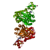 4mv1C 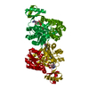 4mv3C 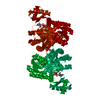 4mv4C 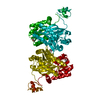 4mv6C  4mv7C 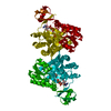 4mv8C  4mv9C 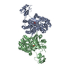 1dv1S S: Starting model for refinement C: citing same article ( |
|---|---|
| Similar structure data |


-Links
 PDBj
PDBj


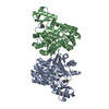
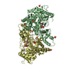
- Assembly
| Deposited unit | 
| ||||||||
|---|---|---|---|---|---|---|---|---|---|
| 1 | 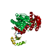
| ||||||||
| Unit cell |
| ||||||||
| Components on special symmetry positions |
|
-Components
| #1: Protein | / Acetyl-CoA carboxylase subunit A / ACC Mass: 51346.555 Da / Num. of mol.: 1 Source method: isolated from a genetically manipulated source Source: (gene. exp.) Haemophilus influenzae Rd KW20 (bacteria)Strain: ATCC 51907 / DSM 11121 / KW20 / Rd / Gene: accC, HI_0972 / Production host: Escherichia coli (E. coli)References: UniProt: P43873, biotin carboxylase, acetyl-CoA carboxylase |
|---|---|
| #2: Chemical | ChemComp-Y7Y /   Mass: 316.373 Da / Num. of mol.: 1 / Source method: obtained synthetically / Formula: C13H20N2O5S Mass: 316.373 Da / Num. of mol.: 1 / Source method: obtained synthetically / Formula: C13H20N2O5S |
| #3: Chemical | ChemComp-ADP / Adenosine diphosphate  Mass: 427.201 Da / Num. of mol.: 1 / Source method: obtained synthetically / Formula: C10H15N5O10P2 / Comment: ADP, energy-carrying molecule*YM Mass: 427.201 Da / Num. of mol.: 1 / Source method: obtained synthetically / Formula: C10H15N5O10P2 / Comment: ADP, energy-carrying molecule*YM |
| #4: Water | ChemComp-HOH / Water Mass: 18.015 Da / Num. of mol.: 84 / Source method: isolated from a natural source / Formula: H2O Mass: 18.015 Da / Num. of mol.: 84 / Source method: isolated from a natural source / Formula: H2O |
-Experimental details
-Experiment
| Experiment | Method: X-RAY DIFFRACTION / Number of used crystals: 1 |
|---|
- Sample preparation
| Crystal | Density Matthews: 2.14 Å3/Da / Density % sol: 42.47 % |
|---|---|
| Crystal grow | Temperature: 298.15 K / Method: vapor diffusion, sitting drop Details: 16% PEG3350 0.2M KCl Protein was incubated with solid N1 -methoxycarbonylbiotin methyl ester for 5 days prior to crystallization, VAPOR DIFFUSION, SITTING DROP, temperature 298.15K |
-Data collection
| Diffraction | Mean temperature: 100 K |
|---|---|
| Diffraction source | Source: SYNCHROTRON / Site: APS  / Beamline: 24-ID-E / Wavelength: 0.97918 Å / Beamline: 24-ID-E / Wavelength: 0.97918 Å |
| Detector | Type: ADSC QUANTUM 315 / Detector: CCD / Date: Oct 31, 2013 / Details: KB MIRRORS |
| Radiation | Monochromator: CRYOGENICALLY-COOLED SINGLE CRYSTAL SI(220) SIDE BOUNCE Protocol: SINGLE WAVELENGTH / Monochromatic (M) / Laue (L): M / Scattering type: x-ray |
| Radiation wavelength | Wavelength: 0.97918 Å / Relative weight: 1 |
| Reflection | Resolution: 1.98→39.72 Å / Num. all: 30073 / Num. obs: 30073 / % possible obs: 99.9 % / Redundancy: 5.6 % / Biso Wilson estimate: 27.7 Å2 / Rsym value: 0.056 / Net I/σ(I): 19.5 |
| Reflection shell | Resolution: 1.98→2.09 Å / Redundancy: 5.7 % / Mean I/σ(I) obs: 2.8 / Num. unique all: 4385 / Rsym value: 0.504 / % possible all: 100 |
- Processing
| Software |
| ||||||||||||||||||||||||||||||||||||||||||||||||||||||||||||||||||||||||||||||||||||||||||||||||||||||||||||||||||||||||||||||||||||||||||||||||||||||||||||||||||||||||||||||||||||||
|---|---|---|---|---|---|---|---|---|---|---|---|---|---|---|---|---|---|---|---|---|---|---|---|---|---|---|---|---|---|---|---|---|---|---|---|---|---|---|---|---|---|---|---|---|---|---|---|---|---|---|---|---|---|---|---|---|---|---|---|---|---|---|---|---|---|---|---|---|---|---|---|---|---|---|---|---|---|---|---|---|---|---|---|---|---|---|---|---|---|---|---|---|---|---|---|---|---|---|---|---|---|---|---|---|---|---|---|---|---|---|---|---|---|---|---|---|---|---|---|---|---|---|---|---|---|---|---|---|---|---|---|---|---|---|---|---|---|---|---|---|---|---|---|---|---|---|---|---|---|---|---|---|---|---|---|---|---|---|---|---|---|---|---|---|---|---|---|---|---|---|---|---|---|---|---|---|---|---|---|---|---|---|---|
| Refinement | Method to determine structure: MOLECULAR REPLACEMENT Starting model: 1DV1 Resolution: 1.98→39.72 Å / Cor.coef. Fo:Fc: 0.962 / Cor.coef. Fo:Fc free: 0.944 / SU B: 8.307 / SU ML: 0.116 / Isotropic thermal model: isotropic / Cross valid method: THROUGHOUT / ESU R: 0.168 / ESU R Free: 0.15 / Stereochemistry target values: MAXIMUM LIKELIHOOD / Details: HYDROGENS HAVE BEEN ADDED IN THE RIDING POSITIONS
| ||||||||||||||||||||||||||||||||||||||||||||||||||||||||||||||||||||||||||||||||||||||||||||||||||||||||||||||||||||||||||||||||||||||||||||||||||||||||||||||||||||||||||||||||||||||
| Solvent computation | Ion probe radii: 0.8 Å / Shrinkage radii: 0.8 Å / VDW probe radii: 1.2 Å / Solvent model: MASK | ||||||||||||||||||||||||||||||||||||||||||||||||||||||||||||||||||||||||||||||||||||||||||||||||||||||||||||||||||||||||||||||||||||||||||||||||||||||||||||||||||||||||||||||||||||||
| Displacement parameters | Biso mean: 44.666 Å2
| ||||||||||||||||||||||||||||||||||||||||||||||||||||||||||||||||||||||||||||||||||||||||||||||||||||||||||||||||||||||||||||||||||||||||||||||||||||||||||||||||||||||||||||||||||||||
| Refinement step | Cycle: LAST / Resolution: 1.98→39.72 Å
| ||||||||||||||||||||||||||||||||||||||||||||||||||||||||||||||||||||||||||||||||||||||||||||||||||||||||||||||||||||||||||||||||||||||||||||||||||||||||||||||||||||||||||||||||||||||
| Refine LS restraints |
|