 Movie
Movie Controller

[English] 日本語
 Yorodumi
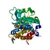
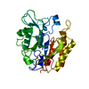
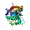
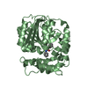
- PDB-4rv3: Crystal structure of a pentafluoro-Phe incorporated Phosphatidyli... -
+ Open data

- Basic information
| Entry | Database: PDB / ID: 4rv3 | ||||||
|---|---|---|---|---|---|---|---|
| Title | Crystal structure of a pentafluoro-Phe incorporated Phosphatidylinositol-specific phospholipase C (H258X)from Staphylococcus aureus | ||||||
 Components Components | 1-phosphatidylinositol phosphodiesterase | ||||||
 Keywords Keywords |  LYASE / cation-pi / TIM barrel / encoded unnatural amino acid phospholipase LYASE / cation-pi / TIM barrel / encoded unnatural amino acid phospholipase | ||||||
| Function / homology |  Function and homology informationphosphatidylinositol diacylglycerol-lyase / phosphatidylinositol diacylglycerol-lyase activity / phosphoric diester hydrolase activity / lipid catabolic process / extracellular region Function and homology informationphosphatidylinositol diacylglycerol-lyase / phosphatidylinositol diacylglycerol-lyase activity / phosphoric diester hydrolase activity / lipid catabolic process / extracellular regionSimilarity search - Function | ||||||
| Biological species |   Staphylococcus aureus (bacteria) Staphylococcus aureus (bacteria) | ||||||
| Method | X-RAY DIFFRACTION / MOLECULAR REPLACEMENT / Resolution: 2 Å | ||||||
 Authors Authors | He, T. / Gershenson, A. / Eyles, S.J. / Gao, J. / Roberts, M.F. | ||||||
 Citation Citation | Journal: J.Biol.Chem. / Year: 2015 Title: Fluorinated Aromatic Amino Acids Distinguish Cation-pi Interactions from Membrane Insertion. Authors: He, T. / Gershenson, A. / Eyles, S.J. / Lee, Y.J. / Liu, W.R. / Wang, J. / Gao, J. / Roberts, M.F. | ||||||
| History |
|
- Structure visualization
| Structure viewer | Molecule: MolmilJmol/JSmol |
|---|
- Downloads & links
-Download
| PDBx/mmCIF format | 4rv3.cif.gz | 72.5 KB | Display | PDBx/mmCIF format |
|---|---|---|---|---|
| PDB format | pdb4rv3.ent.gz | 57.3 KB | Display | PDB format |
| PDBx/mmJSON format | 4rv3.json.gz | Tree view | PDBx/mmJSON format | |
| Others |  Other downloads Other downloads |
-Validation report
| Arichive directory | https://data.pdbj.org/pub/pdb/validation_reports/rv/4rv3ftp://data.pdbj.org/pub/pdb/validation_reports/rv/4rv3 | HTTPS FTP |
|---|
-Related structure data


-Links
 PDBj
PDBj- Assembly
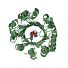
| Deposited unit | 
| ||||||||
|---|---|---|---|---|---|---|---|---|---|
| 1 |
| ||||||||
| Unit cell |
|
-Components
| #1: Protein | Mass: 34296.055 Da / Num. of mol.: 1 / Mutation: H258(PF5) Source method: isolated from a genetically manipulated source Source: (gene. exp.) Staphylococcus aureus (bacteria) / Strain: Newman / Production host: Escherichia coli (E. coli) / Strain (production host): BL21(DE3)References: UniProt: P45723, phosphatidylinositol diacylglycerol-lyase |
|---|---|
| #2: Chemical | ChemComp-ACT / Acetate  Mass: 59.044 Da / Num. of mol.: 1 / Source method: obtained synthetically / Formula: C2H3O2 Mass: 59.044 Da / Num. of mol.: 1 / Source method: obtained synthetically / Formula: C2H3O2 |
| #3: Chemical | ChemComp-INS / Inositol  Mass: 180.156 Da / Num. of mol.: 1 / Source method: obtained synthetically / Formula: C6H12O6 / Comment: neurotransmitter, hormone*YM Mass: 180.156 Da / Num. of mol.: 1 / Source method: obtained synthetically / Formula: C6H12O6 / Comment: neurotransmitter, hormone*YM |
| #4: Water | ChemComp-HOH / Water Mass: 18.015 Da / Num. of mol.: 161 / Source method: isolated from a natural source / Formula: H2O Mass: 18.015 Da / Num. of mol.: 161 / Source method: isolated from a natural source / Formula: H2O |
-Experimental details
-Experiment
| Experiment | Method: X-RAY DIFFRACTION / Number of used crystals: 1 |
|---|
- Sample preparation
| Crystal | Density Matthews: 2.36 Å3/Da / Density % sol: 47.78 % |
|---|---|
| Crystal grow | Temperature: 277 K / Method: vapor diffusion, hanging drop / pH: 4.6 Details: 26% PEG 4000,0.150M ammonium acetate,0.1M sodium acetate, 0.1M magnesium nitrate, pH 4.6, VAPOR DIFFUSION, HANGING DROP, temperature 277K |
-Data collection
| Diffraction | Mean temperature: 120 K |
|---|---|
| Diffraction source | Source: ROTATING ANODE / Type: RIGAKU MICROMAX-007 HF / Wavelength: 1.54 Å |
| Detector | Type: RIGAKU RAXIS IV++ / Detector: IMAGE PLATE / Date: Jan 28, 2014 |
| Radiation | Monochromator: Osmic VariMax / Protocol: SINGLE WAVELENGTH / Monochromatic (M) / Laue (L): M / Scattering type: x-ray |
| Radiation wavelength | Wavelength: 1.54 Å / Relative weight: 1 |
| Reflection | Resolution: 2→49.87 Å / Num. all: 23089 / Num. obs: 22311 / % possible obs: 96.61 % / Observed criterion σ(F): 0 / Observed criterion σ(I): -3 / Redundancy: 4.31 % |
| Reflection shell | Resolution: 2→2.07 Å / Redundancy: 2.13 % / Rmerge(I) obs: 0.199 / Mean I/σ(I) obs: 3.9 / % possible all: 74.6 |
- Processing
| Software |
| |||||||||||||||||||||||||||||||||||||||||||||||||||||||||||||||
|---|---|---|---|---|---|---|---|---|---|---|---|---|---|---|---|---|---|---|---|---|---|---|---|---|---|---|---|---|---|---|---|---|---|---|---|---|---|---|---|---|---|---|---|---|---|---|---|---|---|---|---|---|---|---|---|---|---|---|---|---|---|---|---|---|
| Refinement | Method to determine structure: MOLECULAR REPLACEMENT / Resolution: 2→49.87 Å / SU ML: 0.22 / σ(F): 1.35 / Phase error: 23.12 / Stereochemistry target values: ML
| |||||||||||||||||||||||||||||||||||||||||||||||||||||||||||||||
| Solvent computation | Shrinkage radii: 0.9 Å / VDW probe radii: 1.11 Å / Solvent model: FLAT BULK SOLVENT MODEL | |||||||||||||||||||||||||||||||||||||||||||||||||||||||||||||||
| Refinement step | Cycle: LAST / Resolution: 2→49.87 Å
| |||||||||||||||||||||||||||||||||||||||||||||||||||||||||||||||
| Refine LS restraints |
| |||||||||||||||||||||||||||||||||||||||||||||||||||||||||||||||
| LS refinement shell |
|
