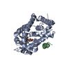
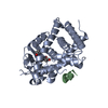
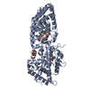
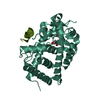
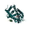
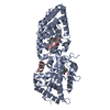
Entry Database : PDB / ID : 4rujTitle Crystal structure of zVDR L337H mutant-VD complex Nuclear receptor coactivator 1 Vitamin D3 receptor A Keywords / / / / / / / / Function / homology Function Domain/homology Component
/ / / / / / / / / / / / / / / / / / / / / / / / / / / / / / / / / / / / / / / / / / / / / / / / / / / / / / / / / / / / / / / / / / / / / / / / / / / / / / / / / / / / / / / / / / / / / / / / / / / / / / / / / / / / / / / / / / / / / / / / / / / / / / / / / / / / / / / / / / / Biological species Danio rerio (zebrafish)Homo sapiens (human)Method / / / Resolution : 2.352 Å Authors Huet, T. / Moras, D. / Rochel, N. Journal : Cell Rep / Year : 2015Title : A vitamin D receptor selectively activated by gemini analogs reveals ligand dependent and independent effects.Authors : Huet, T. / Laverny, G. / Ciesielski, F. / Molnar, F. / Ramamoorthy, T.G. / Belorusova, A.Y. / Antony, P. / Potier, N. / Metzger, D. / Moras, D. / Rochel, N. History Deposition Nov 20, 2014 Deposition site / Processing site Revision 1.0 Oct 7, 2015 Provider / Type Revision 1.1 Jul 17, 2019 Group / Data collection / Refinement description / Category / softwareItem / _software.name / _software.versionRevision 1.2 Feb 28, 2024 Group Advisory / Data collection ... Advisory / Data collection / Database references / Derived calculations Category chem_comp_atom / chem_comp_bond ... chem_comp_atom / chem_comp_bond / database_2 / pdbx_unobs_or_zero_occ_atoms / struct_ref_seq_dif / struct_site Item _database_2.pdbx_DOI / _database_2.pdbx_database_accession ... _database_2.pdbx_DOI / _database_2.pdbx_database_accession / _struct_ref_seq_dif.details / _struct_site.pdbx_auth_asym_id / _struct_site.pdbx_auth_comp_id / _struct_site.pdbx_auth_seq_id
Show all Show less
 Movie
Movie Controller


 Open data
 Basic information
 Components
 Keywords
 transcription factor /
  Function and homology information

 Authors
 Citation
 Structure visualization
 Downloads & links
 Other downloads


 PDBj
PDBj


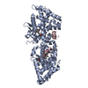
 Assembly
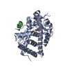


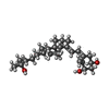
 Mass: 416.636 Da / Num. of mol.: 1 / Source method: obtained synthetically / Formula: C27H44O3
 Mass: 18.015 Da / Num. of mol.: 54 / Source method: isolated from a natural source / Formula: H2O
 Sample preparation
 / Beamline: ID29 / Wavelength: 0.9787 Å
 Processing