 Movie
Movie Controller

[English] 日本語
 Yorodumi
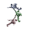
- PDB-4rnf: PaMorA tandem diguanylate cyclase - mutant phosphodiesterase, apo form -
+ Open data

- Basic information
| Entry | Database: PDB / ID: 4rnf | ||||||
|---|---|---|---|---|---|---|---|
| Title | PaMorA tandem diguanylate cyclase - mutant phosphodiesterase, apo form | ||||||
 Components Components | Motility regulator | ||||||
 Keywords Keywords |  Transferase / Hydrolase / Tandem GGDEF and EAL domain / Diguanylate cyclase / Phosphodiesterase / GTP / c-di-GMP Transferase / Hydrolase / Tandem GGDEF and EAL domain / Diguanylate cyclase / Phosphodiesterase / GTP / c-di-GMP | ||||||
| Function / homology |  Function and homology informationnucleotide binding / regulation of DNA-templated transcription / membrane / identical protein binding / metal ion binding Function and homology informationnucleotide binding / regulation of DNA-templated transcription / membrane / identical protein binding / metal ion bindingSimilarity search - Function | ||||||
| Biological species |  Pseudomonas aeruginosa PAO1 (bacteria) Pseudomonas aeruginosa PAO1 (bacteria) | ||||||
| Method | X-RAY DIFFRACTION / SYNCHROTRON / MOLECULAR REPLACEMENT / Resolution: 2.85 Å | ||||||
 Authors Authors | Phippen, C.W. / Tews, I. | ||||||
 Citation Citation | Journal: Febs Lett. / Year: 2014 Title: Formation and dimerization of the phosphodiesterase active site of the Pseudomonas aeruginosa MorA, a bi-functional c-di-GMP regulator. Authors: Phippen, C.W. / Mikolajek, H. / Schlaefli, H.G. / Keevil, C.W. / Webb, J.S. / Tews, I. | ||||||
| History |
|
- Structure visualization
| Structure viewer | Molecule: MolmilJmol/JSmol |
|---|
- Downloads & links
-Download
| PDBx/mmCIF format | 4rnf.cif.gz | 93.7 KB | Display | PDBx/mmCIF format |
|---|---|---|---|---|
| PDB format | pdb4rnf.ent.gz | 70.7 KB | Display | PDB format |
| PDBx/mmJSON format | 4rnf.json.gz | Tree view | PDBx/mmJSON format | |
| Others |  Other downloads Other downloads |
-Validation report
| Arichive directory | https://data.pdbj.org/pub/pdb/validation_reports/rn/4rnfftp://data.pdbj.org/pub/pdb/validation_reports/rn/4rnf | HTTPS FTP |
|---|
-Related structure data


-Links
 PDBj
PDBj


- Assembly
| Deposited unit | 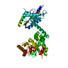
| ||||||||
|---|---|---|---|---|---|---|---|---|---|
| 1 |
| ||||||||
| Unit cell |
| ||||||||
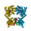
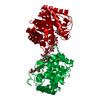
| Details | Active diguanylate cyclase and Active phosphodiesterase are dimers. |
-Components
| #1: Protein | Mass: 50445.535 Da / Num. of mol.: 1 / Fragment: GGDEF domain, EAL domain, UNP residues 978-1409 / Mutation: D1310N, D1311N Source method: isolated from a genetically manipulated source Source: (gene. exp.) Pseudomonas aeruginosa PAO1 (bacteria) / Strain: PA01 / Gene: morA, PA4601 / Plasmid: pET28a / Production host: Escherichia coli (E. coli) / Strain (production host): BL21 (DE3)References: UniProt: Q9HVI8, diguanylate cyclase, cyclic-guanylate-specific phosphodiesterase |
|---|
-Experimental details
-Experiment
| Experiment | Method: X-RAY DIFFRACTION / Number of used crystals: 1 |
|---|
- Sample preparation
| Crystal | Density Matthews: 2.05 Å3/Da / Density % sol: 40.11 % |
|---|---|
| Crystal grow | Temperature: 295 K / pH: 6.5 Details: 0.1M Imidazole; MES, 0.12M 1,6-Hexanediol; 1-Butanol 1,2-Propanediol (racemic); 2-Propanol; 1,4-Butanediol; 1,3-Propanediol, 30% Ethylene glycol; PEG 8000, pH 6.5, VAPOR DIFFUSION, SITTING DROP, temperature 295K |
-Data collection
| Diffraction | Mean temperature: 100 K |
|---|---|
| Diffraction source | Source: SYNCHROTRON / Site: Diamond  / Beamline: I03 / Wavelength: 0.97623 / Beamline: I03 / Wavelength: 0.97623 |
| Detector | Type: DECTRIS PILATUS 6M / Detector: PIXEL / Date: Jul 27, 2013 |
| Radiation | Monochromator: DOUBLE CRYSTAL MONOCHROMATOR 28.290M / Protocol: SINGLE WAVELENGTH / Monochromatic (M) / Laue (L): M / Scattering type: x-ray |
| Radiation wavelength | Wavelength: 0.97623 Å / Relative weight: 1 |
| Reflection | Resolution: 2.85→28.46 Å / Num. obs: 9235 / % possible obs: 98.5 % / Redundancy: 2.8 % / Biso Wilson estimate: 36.34 Å2 / Rmerge(I) obs: 0.174 / Net I/σ(I): 7.4 |
| Reflection shell | Resolution: 2.85→3 Å / Redundancy: 2.8 % / Rmerge(I) obs: 0.578 / Mean I/σ(I) obs: 2.4 / % possible all: 98 |
- Processing
| Software |
| ||||||||||||||||||||||||||||
|---|---|---|---|---|---|---|---|---|---|---|---|---|---|---|---|---|---|---|---|---|---|---|---|---|---|---|---|---|---|
| Refinement | Method to determine structure: MOLECULAR REPLACEMENT / Resolution: 2.85→28.46 Å / SU ML: 0.35 / σ(F): 1.97 / Phase error: 30.86 / Stereochemistry target values: MAXIMUM LIKELIHOOD / Details: HYDROGENS HAVE BEEN ADDED IN THE RIDING
| ||||||||||||||||||||||||||||
| Solvent computation | Shrinkage radii: 0.9 Å / VDW probe radii: 1.11 Å / Solvent model: FLAT BULK SOLVENT MODEL | ||||||||||||||||||||||||||||
| Displacement parameters | Biso mean: 28.2 Å2 | ||||||||||||||||||||||||||||
| Refinement step | Cycle: LAST / Resolution: 2.85→28.46 Å
| ||||||||||||||||||||||||||||
| Refine LS restraints |
| ||||||||||||||||||||||||||||
| LS refinement shell |
|
