 Movie
Movie Controller

[English] 日本語
 Yorodumi
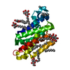
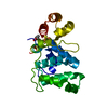
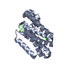
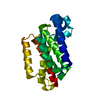
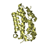
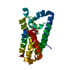
- PDB-2irv: Crystal structure of GlpG, a rhomboid intramembrane serine protease -
+ Open data

- Basic information
| Entry | Database: PDB / ID: 2irv | ||||||
|---|---|---|---|---|---|---|---|
| Title | Crystal structure of GlpG, a rhomboid intramembrane serine protease | ||||||
 Components Components | Protein glpG | ||||||
 Keywords Keywords |  MEMBRANE PROTEIN / cavity / ser-his dyad MEMBRANE PROTEIN / cavity / ser-his dyad | ||||||
| Function / homology |  Function and homology informationrhomboid protease / endopeptidase activity / serine-type endopeptidase activity / proteolysis / identical protein binding / plasma membrane Function and homology informationrhomboid protease / endopeptidase activity / serine-type endopeptidase activity / proteolysis / identical protein binding / plasma membraneSimilarity search - Function | ||||||
| Biological species |  Escherichia coli (E. coli) Escherichia coli (E. coli) | ||||||
| Method | X-RAY DIFFRACTION / SIRAS / Resolution: 2.3 Å | ||||||
 Authors Authors | Bibi, E. / Fass, D. / Ben-Shem, A. | ||||||
 Citation Citation | Journal: Proc.Natl.Acad.Sci.Usa / Year: 2007 Title: Structural basis for intramembrane proteolysis by rhomboid serine proteases. Authors: Ben-Shem, A. / Fass, D. / Bibi, E. | ||||||
| History |
|
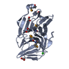
- Structure visualization
| Structure viewer | Molecule: MolmilJmol/JSmol |
|---|
- Downloads & links
-Download
| PDBx/mmCIF format | 2irv.cif.gz | 92.9 KB | Display | PDBx/mmCIF format |
|---|---|---|---|---|
| PDB format | pdb2irv.ent.gz | 69.9 KB | Display | PDB format |
| PDBx/mmJSON format | 2irv.json.gz | Tree view | PDBx/mmJSON format | |
| Others |  Other downloads Other downloads |
-Validation report
| Arichive directory | https://data.pdbj.org/pub/pdb/validation_reports/ir/2irvftp://data.pdbj.org/pub/pdb/validation_reports/ir/2irv | HTTPS FTP |
|---|
-Related structure data
| Related structure data | |
|---|---|
| Similar structure data |


-Links
 PDBj
PDBj
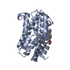
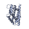
- Assembly
| Deposited unit | 
| ||||||||
|---|---|---|---|---|---|---|---|---|---|
| 1 | 
| ||||||||
| 2 | 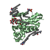
| ||||||||
| Unit cell |
|
-Components
-Protein / Sugars , 2 types, 3 molecules AB
| #1: Protein | Mass: 20556.393 Da / Num. of mol.: 2 / Fragment: protease core, residues 92-273 Source method: isolated from a genetically manipulated source Source: (gene. exp.) Escherichia coli (E. coli) / Strain: K12 / Gene: glpG / Plasmid: pET-28a / Production host: Escherichia coli (E. coli) / Strain (production host): C43(DE3) / References: UniProt: P09391#4: Sugar | ChemComp-LMT / |  Type: D-saccharide / Mass: 510.615 Da / Num. of mol.: 1 / Source method: obtained synthetically / Formula: C24H46O11 / Comment: detergent*YM Type: D-saccharide / Mass: 510.615 Da / Num. of mol.: 1 / Source method: obtained synthetically / Formula: C24H46O11 / Comment: detergent*YM |
|---|
-Non-polymers , 4 types, 102 molecules 


| #2: Chemical | ChemComp-LDA / Lauryldimethylamine oxide Mass: 229.402 Da / Num. of mol.: 16 / Source method: obtained synthetically / Formula: C14H31NO / Comment: LDAO, detergent*YM Mass: 229.402 Da / Num. of mol.: 16 / Source method: obtained synthetically / Formula: C14H31NO / Comment: LDAO, detergent*YM#3: Chemical | ChemComp-PO4 / | Phosphate Mass: 94.971 Da / Num. of mol.: 1 / Source method: obtained synthetically / Formula: PO4 Mass: 94.971 Da / Num. of mol.: 1 / Source method: obtained synthetically / Formula: PO4#5: Chemical | ChemComp-PGV / ( Phosphatidylglycerol Mass: 749.007 Da / Num. of mol.: 4 / Source method: obtained synthetically / Formula: C40H77O10P / Comment: phospholipid*YM Mass: 749.007 Da / Num. of mol.: 4 / Source method: obtained synthetically / Formula: C40H77O10P / Comment: phospholipid*YM#6: Water | ChemComp-HOH / | WaterMass: 18.015 Da / Num. of mol.: 81 / Source method: isolated from a natural source / Formula: H2O |
|---|
-Experimental details
-Experiment
| Experiment | Method: X-RAY DIFFRACTION / Number of used crystals: 1 |
|---|
- Sample preparation
| Crystal | Density Matthews: 2.79 Å3/Da / Density % sol: 55.86 % |
|---|---|
| Crystal grow | Temperature: 277 K / Method: vapor diffusion, hanging drop / pH: 6.5 Details: 100 mM MES, 30% PEG 400, 200 mM CaCl2 in reservoir, pH 6.5, VAPOR DIFFUSION, HANGING DROP, temperature 277K |
-Data collection
| Diffraction | Mean temperature: 120 K |
|---|---|
| Diffraction source | Source: ROTATING ANODE / Type: RIGAKU RU300 / Wavelength: 1.541 Å |
| Detector | Type: RIGAKU RAXIS IV / Detector: IMAGE PLATE / Date: May 1, 2006 |
| Radiation | Protocol: SINGLE WAVELENGTH / Monochromatic (M) / Laue (L): M / Scattering type: x-ray |
| Radiation wavelength | Wavelength: 1.541 Å / Relative weight: 1 |
| Reflection | Resolution: 2.3→50 Å / Num. obs: 17028 / % possible obs: 98.4 % / Observed criterion σ(F): -3 / Redundancy: 3 % / Rsym value: 0.081 / Net I/σ(I): 9.8 |
| Reflection shell | Resolution: 2.3→2.38 Å / Redundancy: 2.8 % / Mean I/σ(I) obs: 1.8 / Rsym value: 0.411 / % possible all: 97.2 |
- Processing
| Software |
| ||||||||||||||||
|---|---|---|---|---|---|---|---|---|---|---|---|---|---|---|---|---|---|
| Refinement | Method to determine structure: SIRAS / Resolution: 2.3→50 Å / Cross valid method: THROUGHOUT / σ(F): 0
| ||||||||||||||||
| Refinement step | Cycle: LAST / Resolution: 2.3→50 Å
| ||||||||||||||||
| Refine LS restraints |
|
