 Movie
Movie Controller


+ Open data

- Basic information
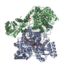
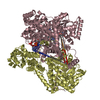
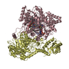
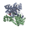
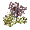
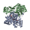
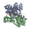
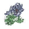
| Entry | Database: PDB / ID: 4req | ||||||
|---|---|---|---|---|---|---|---|
| Title | Methylmalonyl-COA Mutase substrate complex | ||||||
 Components Components | (METHYLMALONYL-COA ... ) x 2 ) x 2 | ||||||
 Keywords Keywords | ISOMERASE / MUTASE / INTRAMOLECULAR TRANSFERASE | ||||||
| Function / homology |  Function and homology information Function and homology informationlactate fermentation to propionate and acetate / propionate metabolic process, methylmalonyl pathway / methylmalonyl-CoA mutase / methylmalonyl-CoA mutase activity / cobalamin binding / metal ion binding / cytoplasmSimilarity search - Function | ||||||
| Biological species |  Propionibacterium freudenreichii subsp. shermanii (bacteria) Propionibacterium freudenreichii subsp. shermanii (bacteria) | ||||||
| Method | X-RAY DIFFRACTION / SYNCHROTRON / MOLECULAR REPLACEMENT / Resolution: 2.2 Å | ||||||
 Authors Authors | Evans, P.R. / Mancia, F. | ||||||
 Citation Citation | Journal: Structure / Year: 1998 Title: Conformational changes on substrate binding to methylmalonyl CoA mutase and new insights into the free radical mechanism. Authors: Mancia, F. / Evans, P.R. #1: Journal: Vitamin B12 and B12-Proteins : Lectures Presented at the 4Th European Symposium on Vitamin B12 and B12-ProteinsYear: 1998 Title: Insights on the Reaction Mechanism of Methylmalonyl-Coa Mutase from the Crystal Structure Authors: Evans, P.R. / Mancia, F. | ||||||
| History |
|
- Structure visualization
| Structure viewer | Molecule: MolmilJmol/JSmol |
|---|
- Downloads & links
-Download
| PDBx/mmCIF format | 4req.cif.gz | 572 KB | Display | PDBx/mmCIF format |
|---|---|---|---|---|
| PDB format | pdb4req.ent.gz | 453.8 KB | Display | PDB format |
| PDBx/mmJSON format | 4req.json.gz | Tree view | PDBx/mmJSON format | |
| Others |  Other downloads Other downloads |
-Validation report
| Arichive directory | https://data.pdbj.org/pub/pdb/validation_reports/re/4reqftp://data.pdbj.org/pub/pdb/validation_reports/re/4req | HTTPS FTP |
|---|
-Related structure data
| Related structure data | 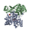 2reqC  3reqC  1reqS S: Starting model for refinement C: citing same article ( |
|---|---|
| Similar structure data |

-Links
 PDBj
PDBj- Assembly
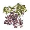
| Deposited unit | 
| ||||||||
|---|---|---|---|---|---|---|---|---|---|
| 1 | 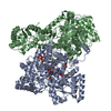
| ||||||||
| 2 | 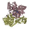
| ||||||||
| Unit cell |
| ||||||||
| Noncrystallographic symmetry (NCS) | NCS oper: (Code: given Matrix: (-0.462866, -0.001054, 0.886428), Vector : Details | THE ASYMMETRIC UNIT OF THE CRYSTAL CONTAINS TWO HETERODIMERIC MOLECULES, EACH WITH AN ALPHA CHAIN (CHAINS A AND C, CORRESPONDING TO GENE MUTB) AND A BETA CHAIN (CHAINS B AND D, CORRESPONDING TO GENE MUTA). MOLECULE 1 CONSISTS OF CHAINS A (ALPHA), B (BETA), WITH GLYCEROL CHAIN G AND WATERS X. MOLECULE 2 CONSISTS OF CHAINS C (ALPHA), D (BETA), WITH GLYCEROL CHAIN H AND WATERS Y. CHAINS A AND C INCLUDE COENZYME B12 (RESIDUE 800), A 1:1 MIXTURE OF SUBSTRATE AND PRODUCT, SUCCINYL-COA (RESIDUE 801), METHYLMALONYL-COA (RESIDUE 802), AND A HALF-OCCUPIED 5'-DEOXYADENOSINE INTERMEDIATE. | |
-Components
-METHYLMALONYL-COA ... , 2 types, 4 molecules ACBD
| #1: Protein | Mass: 80137.852 Da / Num. of mol.: 2 / Source method: isolated from a natural source Details: THE 2 GENES MUTA (BETA CHAIN) AND MUTB (ALPHA CHAIN) ARE COEXPRESSED FROM THE SAME PLASMID Source: (natural) Propionibacterium freudenreichii subsp. shermanii (bacteria)Plasmid: PMEX1 / Species: Propionibacterium freudenreichii / Strain: NCIB 9885 / References: UniProt: P11653, methylmalonyl-CoA mutase#2: Protein | Mass: 69430.188 Da / Num. of mol.: 2 / Source method: isolated from a natural source Details: THE 2 GENES MUTA (BETA CHAIN) AND MUTB (ALPHA CHAIN) ARE COEXPRESSED FROM THE SAME PLASMID Source: (natural) Propionibacterium freudenreichii subsp. shermanii (bacteria)Plasmid: PMEX1 / Species: Propionibacterium freudenreichii / Strain: NCIB 9885 / References: UniProt: P11652, methylmalonyl-CoA mutase |
|---|
-Non-polymers , 6 types, 1298 molecules 


| #3: Chemical | Vitamin B12 Mass: 1330.356 Da / Num. of mol.: 2 / Source method: obtained synthetically / Formula: C62H89CoN13O14P Mass: 1330.356 Da / Num. of mol.: 2 / Source method: obtained synthetically / Formula: C62H89CoN13O14P#4: Chemical | Succinyl-CoA Mass: 867.607 Da / Num. of mol.: 2 / Source method: obtained synthetically / Formula: C25H40N7O19P3S Mass: 867.607 Da / Num. of mol.: 2 / Source method: obtained synthetically / Formula: C25H40N7O19P3S#5: Chemical | Methylmalonyl-CoA Mass: 867.607 Da / Num. of mol.: 2 / Source method: obtained synthetically / Formula: C25H40N7O19P3S Mass: 867.607 Da / Num. of mol.: 2 / Source method: obtained synthetically / Formula: C25H40N7O19P3S#6: Chemical | Deoxyadenosine Mass: 251.242 Da / Num. of mol.: 2 / Source method: obtained synthetically / Formula: C10H13N5O3 Mass: 251.242 Da / Num. of mol.: 2 / Source method: obtained synthetically / Formula: C10H13N5O3#7: Chemical | ChemComp-GOL / Glycerol Mass: 92.094 Da / Num. of mol.: 4 / Source method: obtained synthetically / Formula: C3H8O3 Mass: 92.094 Da / Num. of mol.: 4 / Source method: obtained synthetically / Formula: C3H8O3#8: Water | ChemComp-HOH / | WaterMass: 18.015 Da / Num. of mol.: 1286 / Source method: isolated from a natural source / Formula: H2O |
|---|
-Details
| Nonpolymer details | THE COBALAMIN HAS BEEN MODELLED AS THE 5-COORDINATES REDUCED COB(II)ALAMIN (B12R), SINCE THERE IS ...THE COBALAMIN HAS BEEN MODELLED AS THE 5-COORDINATE |
|---|
-Experimental details
-Experiment
| Experiment | Method: X-RAY DIFFRACTION / Number of used crystals: 1 |
|---|
- Sample preparation
| Crystal | Density Matthews: 2.74 Å3/Da / Density % sol: 48 % |
|---|---|
| Crystal grow | pH: 7.5 / Details: pH 7.5 |
-Data collection
| Diffraction | Mean temperature: 95 K |
|---|---|
| Diffraction source | Source: SYNCHROTRON / Site: ELETTRA  / Beamline: 5.2R / Wavelength: 1.24 / Beamline: 5.2R / Wavelength: 1.24 |
| Detector | Type: MARRESEARCH / Detector: IMAGE PLATE AREA DETECTOR / Date: Sep 1, 1995 / Details: MIRROR |
| Radiation | Monochromator: SI(111) / Monochromatic (M) / Laue (L): M / Scattering type: x-ray |
| Radiation wavelength | Wavelength: 1.24 Å / Relative weight: 1 |
| Reflection | Resolution: 2.2→20 Å / Num. obs: 164181 / % possible obs: 99.8 % / Observed criterion σ(I): 3.5 / Redundancy: 4.3 % / Biso Wilson estimate: 40 Å2 / Rmerge(I) obs: 0.103 / Rsym value: 0.103 / Net I/σ(I): 5.3 |
| Reflection shell | Resolution: 2.2→2.32 Å / Redundancy: 3.5 % / Rmerge(I) obs: 0.379 / Mean I/σ(I) obs: 1.6 / Rsym value: 0.379 / % possible all: 99.9 |
- Processing
| Software |
| ||||||||||||||||||||||||||||||||||||||||||||||||||||||||||||||||||||||||||||||||||||
|---|---|---|---|---|---|---|---|---|---|---|---|---|---|---|---|---|---|---|---|---|---|---|---|---|---|---|---|---|---|---|---|---|---|---|---|---|---|---|---|---|---|---|---|---|---|---|---|---|---|---|---|---|---|---|---|---|---|---|---|---|---|---|---|---|---|---|---|---|---|---|---|---|---|---|---|---|---|---|---|---|---|---|---|---|---|
| Refinement | Method to determine structure: MOLECULAR REPLACEMENT Starting model: PDB ENTRY 1REQ Resolution: 2.2→20 Å / Cross valid method: THROUGHOUT / σ(F): 0
| ||||||||||||||||||||||||||||||||||||||||||||||||||||||||||||||||||||||||||||||||||||
| Displacement parameters | Biso mean: 38 Å2 | ||||||||||||||||||||||||||||||||||||||||||||||||||||||||||||||||||||||||||||||||||||
| Refine analyze | Luzzati coordinate error obs: 0.29 Å | ||||||||||||||||||||||||||||||||||||||||||||||||||||||||||||||||||||||||||||||||||||
| Refinement step | Cycle: LAST / Resolution: 2.2→20 Å
| ||||||||||||||||||||||||||||||||||||||||||||||||||||||||||||||||||||||||||||||||||||
| Refine LS restraints |
|
