 Movie
Movie Controller

[English] 日本語
 Yorodumi
- PDB-4rbx: Crystal structure of human alpha-defensin 5, HD5 (Glu21Arg mutant) -
+ Open data

- Basic information
| Entry | Database: PDB / ID: 4rbx | ||||||
|---|---|---|---|---|---|---|---|
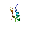
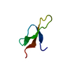
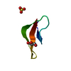
| Title | Crystal structure of human alpha-defensin 5, HD5 (Glu21Arg mutant) | ||||||
 Components Components | Defensin-5 | ||||||
 Keywords Keywords | ANTIMICROBIAL PROTEIN / MUTANT E21R-HD5 / BETA-SHEET / ANTIMICROBIAL PEPTIDE / PANETH CELLS DEFENSIN / HUMAN ALPHA-DEFENSIN | ||||||
| Function / homology |  Function and homology information Function and homology informationpositive regulation of membrane permeability / Defensins / disruption of plasma membrane integrity in another organism / killing by host of symbiont cells / Alpha-defensins / defense response to fungus / transport vesicle / innate immune response in mucosa / secretory granule / positive regulation of interleukin-8 production ...positive regulation of membrane permeability / Defensins / disruption of plasma membrane integrity in another organism / killing by host of symbiont cells / Alpha-defensins / defense response to fungus / transport vesicle / innate immune response in mucosa / secretory granule / positive regulation of interleukin-8 production / Golgi lumen / antimicrobial humoral immune response mediated by antimicrobial peptide / midbody / antibacterial humoral response / protein homotetramerization / secretory granule lumen / killing of cells of another organism / cellular response to lipopolysaccharide / defense response to Gram-negative bacterium / defense response to Gram-positive bacterium / intracellular membrane-bounded organelle / innate immune response / protein homodimerization activity / extracellular space / extracellular regionSimilarity search - Function | ||||||
| Biological species |  Homo sapiens (human) Homo sapiens (human) | ||||||
| Method | X-RAY DIFFRACTION / SYNCHROTRON / MOLECULAR REPLACEMENT / Resolution: 1.1 Å | ||||||
 Authors Authors | Pazgier, M. / Gohain, N. / Tolbert, W.D. | ||||||
 Citation Citation | Journal: J.Med.Chem. / Year: 2015 Title: Design of a potent antibiotic peptide based on the active region of human defensin 5. Authors: Wang, C. / Shen, M. / Gohain, N. / Tolbert, W.D. / Chen, F. / Zhang, N. / Yang, K. / Wang, A. / Su, Y. / Cheng, T. / Zhao, J. / Pazgier, M. / Wang, J. | ||||||
| History |
|
- Structure visualization
| Structure viewer | Molecule: MolmilJmol/JSmol |
|---|
- Downloads & links
-Download
| PDBx/mmCIF format | 4rbx.cif.gz | 25.4 KB | Display | PDBx/mmCIF format |
|---|---|---|---|---|
| PDB format | pdb4rbx.ent.gz | 18.3 KB | Display | PDB format |
| PDBx/mmJSON format | 4rbx.json.gz | Tree view | PDBx/mmJSON format | |
| Others |  Other downloads Other downloads |
-Validation report
| Arichive directory | https://data.pdbj.org/pub/pdb/validation_reports/rb/4rbxftp://data.pdbj.org/pub/pdb/validation_reports/rb/4rbx | HTTPS FTP |
|---|
-Related structure data
| Related structure data | 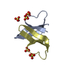 4rbwC  1zmpS S: Starting model for refinement C: citing same article ( |
|---|---|
| Similar structure data |


-Links
 PDBj
PDBj


- Assembly
| Deposited unit | 
| ||||||||
|---|---|---|---|---|---|---|---|---|---|
| 1 |
| ||||||||
| Unit cell |
|
-Components
| #1: Protein/peptide | / HD5(63-94) Mass: 3622.308 Da / Num. of mol.: 1 / Mutation: E21R / Source method: obtained synthetically / Source: (synth.) Homo sapiens (human) / References: UniProt: Q01523 | ||
|---|---|---|---|
| #2: Chemical | Sulfate  Mass: 96.063 Da / Num. of mol.: 3 / Source method: obtained synthetically / Formula: SO4 Mass: 96.063 Da / Num. of mol.: 3 / Source method: obtained synthetically / Formula: SO4#3: Water | ChemComp-HOH / | Water Mass: 18.015 Da / Num. of mol.: 34 / Source method: isolated from a natural source / Formula: H2O Mass: 18.015 Da / Num. of mol.: 34 / Source method: isolated from a natural source / Formula: H2O |
-Experimental details
-Experiment
| Experiment | Method: X-RAY DIFFRACTION / Number of used crystals: 1 |
|---|
- Sample preparation
| Crystal | Density Matthews: 2.39 Å3/Da / Density % sol: 48.45 % |
|---|---|
| Crystal grow | Temperature: 298 K / Method: vapor diffusion, hanging drop / pH: 7.5 Details: 6.6% PEG 8000, 3.3% Isopropanol, 0.2M Ammonium sulfate and 0.1M Hepes pH7.5, VAPOR DIFFUSION, HANGING DROP, temperature 298K |
-Data collection
| Diffraction | Mean temperature: 100 K |
|---|---|
| Diffraction source | Source: SYNCHROTRON / Site: SSRL  / Beamline: BL12-2 / Wavelength: 0.979 Å / Beamline: BL12-2 / Wavelength: 0.979 Å |
| Detector | Type: PSI PILATUS 6M / Detector: PIXEL / Date: Jan 31, 2014 |
| Radiation | Monochromator: Liquid nitrogen-cooled double crystal / Protocol: SINGLE WAVELENGTH / Monochromatic (M) / Laue (L): M / Scattering type: x-ray |
| Radiation wavelength | Wavelength: 0.979 Å / Relative weight: 1 |
| Reflection | Resolution: 1.1→38.4 Å / Num. all: 19818 / Num. obs: 18233 / % possible obs: 92.4 % / Observed criterion σ(F): 0 / Observed criterion σ(I): 0 / Redundancy: 28.3 % / Rmerge(I) obs: 0.07 / Net I/σ(I): 34.6 |
| Reflection shell | Resolution: 1.1→1.13 Å / Redundancy: 11.5 % / Rmerge(I) obs: 0.382 / Mean I/σ(I) obs: 4.5 / Num. unique all: 897 / % possible all: 98.2 |
- Processing
| Software |
| |||||||||||||||||||||||||||||||||||||||||||||||||||||||||||||||||||||||||||||
|---|---|---|---|---|---|---|---|---|---|---|---|---|---|---|---|---|---|---|---|---|---|---|---|---|---|---|---|---|---|---|---|---|---|---|---|---|---|---|---|---|---|---|---|---|---|---|---|---|---|---|---|---|---|---|---|---|---|---|---|---|---|---|---|---|---|---|---|---|---|---|---|---|---|---|---|---|---|---|
| Refinement | Method to determine structure: MOLECULAR REPLACEMENT Starting model: PDB entry 1ZMP Resolution: 1.1→36.336 Å / SU ML: 0.1 / Cross valid method: THROUGHOUT / σ(F): 1.38 / Phase error: 17.51 / Stereochemistry target values: Engh & Huber
| |||||||||||||||||||||||||||||||||||||||||||||||||||||||||||||||||||||||||||||
| Solvent computation | Shrinkage radii: 0.9 Å / VDW probe radii: 1.11 Å / Solvent model: FLAT BULK SOLVENT MODEL | |||||||||||||||||||||||||||||||||||||||||||||||||||||||||||||||||||||||||||||
| Refinement step | Cycle: LAST / Resolution: 1.1→36.336 Å
| |||||||||||||||||||||||||||||||||||||||||||||||||||||||||||||||||||||||||||||
| Refine LS restraints |
| |||||||||||||||||||||||||||||||||||||||||||||||||||||||||||||||||||||||||||||
| LS refinement shell |
|
