 Movie
Movie Controller

[English] 日本語
 Yorodumi
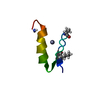
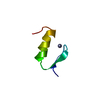
- PDB-3pis: Crystal Structure of Carcinoscorpius rotundicauda Serine Protease... -
+ Open data

- Basic information
| Entry | Database: PDB / ID: 3pis | ||||||
|---|---|---|---|---|---|---|---|
| Title | Crystal Structure of Carcinoscorpius rotundicauda Serine Protease Inhibitor Domain 1 | ||||||
 Components Components | Kazal-type serine protease inhibitor SPI-1 | ||||||
 Keywords Keywords | HYDROLASE INHIBITOR / typical non-classical Kazal type inhibitor Fold / serine protease inhibitors (uncharacterized) | ||||||
| Function / homology |  Kazal-type serine protease inhibitor domain / Kazal type serine protease inhibitors / Kazal domain superfamily / Kazal domain / Kazal domain profile. / Kazal-type serine protease inhibitor SPI-1 Kazal-type serine protease inhibitor domain / Kazal type serine protease inhibitors / Kazal domain superfamily / Kazal domain / Kazal domain profile. / Kazal-type serine protease inhibitor SPI-1 Function and homology information Function and homology information | ||||||
| Biological species |  Carcinoscorpius rotundicauda (Southeast Asian horseshoe crab) Carcinoscorpius rotundicauda (Southeast Asian horseshoe crab) | ||||||
| Method | X-RAY DIFFRACTION / MIR / Resolution: 2 Å | ||||||
 Authors Authors | Giri, P.K. / Tang, X.H. / Sivaraman, J. | ||||||
 Citation Citation | Journal: To be Published Title: Modifying the Substrate Specificity of Carcinoscorpius rotundicauda Serine Protease Inhibitor Domain 1 to Target Thrombin Authors: Giri, P.K. / Tang, X.H. / Thangamani, S. / Shenoy, R.T. / Ding, J.L. / Swaminathan, K. / Sivaraman, J. | ||||||
| History |
|
- Structure visualization
| Structure viewer | Molecule: MolmilJmol/JSmol |
|---|
- Downloads & links
-Download
| PDBx/mmCIF format | 3pis.cif.gz | 22.9 KB | Display | PDBx/mmCIF format |
|---|---|---|---|---|
| PDB format | pdb3pis.ent.gz | 17.2 KB | Display | PDB format |
| PDBx/mmJSON format | 3pis.json.gz | Tree view | PDBx/mmJSON format | |
| Others |  Other downloads Other downloads |
-Validation report
| Arichive directory | https://data.pdbj.org/pub/pdb/validation_reports/pi/3pisftp://data.pdbj.org/pub/pdb/validation_reports/pi/3pis | HTTPS FTP |
|---|
-Related structure data
| Similar structure data |
|---|


-Links
 PDBj
PDBj
- Assembly
| Deposited unit | 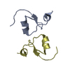
| ||||||||
|---|---|---|---|---|---|---|---|---|---|
| 1 | 
| ||||||||
| 2 | 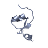
| ||||||||
| Unit cell |
|
-Components
| #1: Protein/peptide | Mass: 4605.021 Da / Num. of mol.: 2 / Fragment: UNP RESIDUES 26-65 Source method: isolated from a genetically manipulated source Source: (gene. exp.) Carcinoscorpius rotundicauda (Southeast Asian horseshoe crab)Gene: SPI-1 / Production host:  Escherichia coli (E. coli) / Strain (production host): BL21 / References: UniProt: A1X1V8 Escherichia coli (E. coli) / Strain (production host): BL21 / References: UniProt: A1X1V8#2: Water | ChemComp-HOH / | Water Mass: 18.015 Da / Num. of mol.: 55 / Source method: isolated from a natural source / Formula: H2O Mass: 18.015 Da / Num. of mol.: 55 / Source method: isolated from a natural source / Formula: H2O |
|---|
-Experimental details
-Experiment
| Experiment | Method: X-RAY DIFFRACTION / Number of used crystals: 1 |
|---|
- Sample preparation
| Crystal | Density Matthews: 1.85 Å3/Da / Density % sol: 33.57 % |
|---|---|
| Crystal grow | Temperature: 293 K / Method: vapor diffusion, hanging drop / pH: 8.5 Details: 0.4M mono ammonium dihydrogen sulphate, 0.1M Tris-HCl pH8.5, VAPOR DIFFUSION, HANGING DROP, temperature 293K |
-Data collection
| Diffraction | Mean temperature: 100 K |
|---|---|
| Diffraction source | Source: ROTATING ANODE / Type: BRUKER AXS MICROSTAR / Wavelength: 1.54 Å |
| Detector | Type: Bruker Platinum 135 / Detector: CCD / Date: Nov 16, 2009 |
| Radiation | Monochromator: SAGITALLY FOCUSED Si(111) / Protocol: SINGLE WAVELENGTH / Monochromatic (M) / Laue (L): M / Scattering type: x-ray |
| Radiation wavelength | Wavelength: 1.54 Å / Relative weight: 1 |
| Reflection | Resolution: 2→50 Å / Num. all: 4535 / Num. obs: 4535 / % possible obs: 97.3 % / Observed criterion σ(F): 1 / Observed criterion σ(I): 1 |
| Reflection shell | Resolution: 2→2.07 Å / Redundancy: 3.2 % / Num. unique all: 381 / % possible all: 80.7 |
- Processing
| Software |
| |||||||||||||||||||||||||
|---|---|---|---|---|---|---|---|---|---|---|---|---|---|---|---|---|---|---|---|---|---|---|---|---|---|---|
| Refinement | Method to determine structure: MIR / Resolution: 2→20 Å / σ(F): 2 / Stereochemistry target values: Engh & Huber
| |||||||||||||||||||||||||
| Refinement step | Cycle: LAST / Resolution: 2→20 Å
| |||||||||||||||||||||||||
| Refine LS restraints |
|
