 Movie
Movie Controller


+ Open data

- Basic information
| Entry | Database: PDB / ID: 4r7z | ||||||
|---|---|---|---|---|---|---|---|
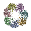
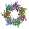
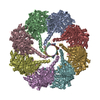
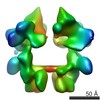
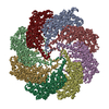
| Title | PfMCM-AAA double-octamer | ||||||
 Components Components | Cell division control protein 21 | ||||||
 Keywords Keywords |  HYDROLASE / AAA+ / MCM / helicase / ATPase / DNA replication HYDROLASE / AAA+ / MCM / helicase / ATPase / DNA replication | ||||||
| Function / homology |  Function and homology information Function and homology informationintein-mediated protein splicing / DNA duplex unwinding / endonuclease activity / DNA helicase / cell division / DNA binding / ATP binding / metal ion bindingSimilarity search - Function | ||||||
| Biological species |   Pyrococcus furiosus (archaea) Pyrococcus furiosus (archaea) | ||||||
| Method | X-RAY DIFFRACTION / SYNCHROTRON / MOLECULAR REPLACEMENT / Resolution: 3.8 Å | ||||||
 Authors Authors | Miller, J.M. / Arachea, B.T. / Epling, L.B. / Enemark, E.J. | ||||||
 Citation Citation | Journal: Elife / Year: 2014 Title: Analysis of the crystal structure of an active MCM hexamer. Authors: Miller, J.M. / Arachea, B.T. / Epling, L.B. / Enemark, E.J. | ||||||
| History |
|
- Structure visualization
| Structure viewer | Molecule: MolmilJmol/JSmol |
|---|
- Downloads & links
-Download
| PDBx/mmCIF format | 4r7z.cif.gz | 867.8 KB | Display | PDBx/mmCIF format |
|---|---|---|---|---|
| PDB format | pdb4r7z.ent.gz | 732.9 KB | Display | PDB format |
| PDBx/mmJSON format | 4r7z.json.gz | Tree view | PDBx/mmJSON format | |
| Others |  Other downloads Other downloads |
-Validation report
| Arichive directory | https://data.pdbj.org/pub/pdb/validation_reports/r7/4r7zftp://data.pdbj.org/pub/pdb/validation_reports/r7/4r7z | HTTPS FTP |
|---|
-Related structure data
| Related structure data |  4r7yC 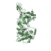 4fdgS C: citing same article ( S: Starting model for refinement |
|---|---|
| Similar structure data |

-Links
 PDBj
PDBj


- Assembly
| Deposited unit | 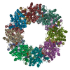
| ||||||||||||||||||||||||||||||||||||||||||||||||||||||||||||||||||||
|---|---|---|---|---|---|---|---|---|---|---|---|---|---|---|---|---|---|---|---|---|---|---|---|---|---|---|---|---|---|---|---|---|---|---|---|---|---|---|---|---|---|---|---|---|---|---|---|---|---|---|---|---|---|---|---|---|---|---|---|---|---|---|---|---|---|---|---|---|---|
| 1 |
| ||||||||||||||||||||||||||||||||||||||||||||||||||||||||||||||||||||
| Unit cell |
| ||||||||||||||||||||||||||||||||||||||||||||||||||||||||||||||||||||
| Noncrystallographic symmetry (NCS) | NCS oper:
|
-Components
| #1: Protein | Mass: 37185.352 Da / Num. of mol.: 16 / Fragment: AAA+ domain of PfMCM (UNP 263-361/729-966) Source method: isolated from a genetically manipulated source Details: Constructed as Sumo fusion (Mossessova and Lima, 2000) Source: (gene. exp.) Pyrococcus furiosus (archaea) / Strain: ATCC 43587 / DSM 3638 / JCM 8422 / Vc1 / Gene: MCM, PF0482 / Plasmid: pRSF-duet / Production host:  Escherichia coli (E. coli) / Strain (production host): BL21(DE3)-RIPL / References: UniProt: Q8U3I4 Escherichia coli (E. coli) / Strain (production host): BL21(DE3)-RIPL / References: UniProt: Q8U3I4#2: Chemical | ChemComp-ADP / Adenosine diphosphate  Mass: 427.201 Da / Num. of mol.: 16 / Source method: obtained synthetically / Formula: C10H15N5O10P2 / Comment: ADP, energy-carrying molecule*YM Mass: 427.201 Da / Num. of mol.: 16 / Source method: obtained synthetically / Formula: C10H15N5O10P2 / Comment: ADP, energy-carrying molecule*YM#3: Chemical | ChemComp-MG /   Mass: 24.305 Da / Num. of mol.: 16 / Source method: obtained synthetically / Formula: Mg Mass: 24.305 Da / Num. of mol.: 16 / Source method: obtained synthetically / Formula: MgSequence details | The UNK region in the sequence is due to unknown registration. The missing sequence is ...The UNK region in the sequence is due to unknown registration. The missing sequence is GKSSSAAGLT | |
|---|
-Experimental details
-Experiment
| Experiment | Method: X-RAY DIFFRACTION / Number of used crystals: 1 |
|---|
- Sample preparation
| Crystal | Density Matthews: 3.09 Å3/Da / Density % sol: 60.21 % |
|---|---|
| Crystal grow | Temperature: 300 K / Method: vapor diffusion, hanging drop / pH: 6 Details: 6 mg/mL PfMCM-AAA; 5 mM ADP; 50 mM MgCl2; 18 mM Hepes, pH 7.6; 180 mM NaCl; 4.5 mM 2-mercaptoethanol mixed 1:1 with 50 mM sodium cacodylate, pH 6.0; 50 mM magnesium acetate; 30% MPD; 5% ...Details: 6 mg/mL PfMCM-AAA; 5 mM ADP; 50 mM MgCl2; 18 mM Hepes, pH 7.6; 180 mM NaCl; 4.5 mM 2-mercaptoethanol mixed 1:1 with 50 mM sodium cacodylate, pH 6.0; 50 mM magnesium acetate; 30% MPD; 5% glycerol, VAPOR DIFFUSION, HANGING DROP, temperature 300K |
-Data collection
| Diffraction | Mean temperature: 100 K |
|---|---|
| Diffraction source | Source: SYNCHROTRON / Site: APS  / Beamline: 22-BM / Wavelength: 1 Å / Beamline: 22-BM / Wavelength: 1 Å |
| Detector | Type: MARMOSAIC 225 mm CCD / Detector: CCD / Date: Jun 8, 2012 |
| Radiation | Monochromator: Si(111) / Protocol: SINGLE WAVELENGTH / Monochromatic (M) / Laue (L): M / Scattering type: x-ray |
| Radiation wavelength | Wavelength: 1 Å / Relative weight: 1 |
| Reflection | Resolution: 3.8→50 Å / Num. all: 70730 / Num. obs: 69970 / % possible obs: 98.9 % / Observed criterion σ(I): -3 / Redundancy: 3.3 % / Rsym value: 0.169 / Net I/σ(I): 8.3 |
| Reflection shell | Resolution: 3.8→3.94 Å / Redundancy: 2.6 % / Mean I/σ(I) obs: 2.41 / Num. unique all: 7090 / Rsym value: 0.429 / % possible all: 97 |
- Processing
| Software |
| ||||||||||||||||||||||||||||||||||||||||||||||||||||||||||||||||||||||||||||||||
|---|---|---|---|---|---|---|---|---|---|---|---|---|---|---|---|---|---|---|---|---|---|---|---|---|---|---|---|---|---|---|---|---|---|---|---|---|---|---|---|---|---|---|---|---|---|---|---|---|---|---|---|---|---|---|---|---|---|---|---|---|---|---|---|---|---|---|---|---|---|---|---|---|---|---|---|---|---|---|---|---|---|
| Refinement | Method to determine structure: MOLECULAR REPLACEMENT Starting model: placement of 16 copies of the ATPase domain of 4FDG (Slaymaker et al., 2013) Resolution: 3.8→48.37 Å / Rfactor Rfree error: 0.005 / Data cutoff high absF: 4895965.68 / Data cutoff low absF: 0 / Isotropic thermal model: RESTRAINED / Cross valid method: THROUGHOUT / σ(F): 0 / Stereochemistry target values: Engh & Huber Details: BULK SOLVENT MODEL USED. Strict NCS was used using the matrices provided and only chain A coordinates
| ||||||||||||||||||||||||||||||||||||||||||||||||||||||||||||||||||||||||||||||||
| Solvent computation | Solvent model: FLAT MODEL / Bsol: 50.0837 Å2 / ksol: 0.3 e/Å3 | ||||||||||||||||||||||||||||||||||||||||||||||||||||||||||||||||||||||||||||||||
| Displacement parameters | Biso mean: 91.2 Å2
| ||||||||||||||||||||||||||||||||||||||||||||||||||||||||||||||||||||||||||||||||
| Refine analyze |
| ||||||||||||||||||||||||||||||||||||||||||||||||||||||||||||||||||||||||||||||||
| Refinement step | Cycle: LAST / Resolution: 3.8→48.37 Å
| ||||||||||||||||||||||||||||||||||||||||||||||||||||||||||||||||||||||||||||||||
| Refine LS restraints |
| ||||||||||||||||||||||||||||||||||||||||||||||||||||||||||||||||||||||||||||||||
| Refine LS restraints NCS | NCS model details: CONSTR | ||||||||||||||||||||||||||||||||||||||||||||||||||||||||||||||||||||||||||||||||
| LS refinement shell | Resolution: 3.8→3.94 Å / Rfactor Rfree error: 0.019 / Total num. of bins used: 10
| ||||||||||||||||||||||||||||||||||||||||||||||||||||||||||||||||||||||||||||||||
| Xplor file |
|
