MCM complex / intein-mediated protein splicing / DNA duplex unwinding / helicase activity / single-stranded DNA binding / endonuclease activity / DNA helicase / DNA replication / cell division / ATP hydrolysis activity ...MCM complex / intein-mediated protein splicing / DNA duplex unwinding / helicase activity / single-stranded DNA binding / endonuclease activity / DNA helicase / DNA replication / cell division / ATP hydrolysis activity / DNA binding / ATP binding / identical protein binding / metal ion binding Similarity search - Function
mini-chromosome maintenance (MCM) complex, chain A, domain 1 / mini-chromosome maintenance (MCM) complex, chain A, domain 1 / : / MCM protein C-terminal winged helix-turn-helix domain / Rubrerythrin, domain 2 - #10 / Intein splicing domain / Intein / Intein DOD homing endonuclease / Intein DOD-type homing endonuclease domain profile. / Intein C-terminal splicing region ...mini-chromosome maintenance (MCM) complex, chain A, domain 1 / mini-chromosome maintenance (MCM) complex, chain A, domain 1 / : / MCM protein C-terminal winged helix-turn-helix domain / Rubrerythrin, domain 2 - #10 / Intein splicing domain / Intein / Intein DOD homing endonuclease / Intein DOD-type homing endonuclease domain profile. / Intein C-terminal splicing region / Intein C-terminal splicing motif profile. / Hint domain C-terminal / Hint (Hedgehog/Intein) domain C-terminal region / MCM N-terminal domain / MCM N-terminal domain / MCM OB domain / MCM OB domain / Intein N-terminal splicing region / Mini-chromosome maintenance protein / MCM, AAA-lid domain / MCM P-loop domain / MCM AAA-lid domain / MCM family domain profile. / Intein N-terminal splicing motif profile. / minichromosome maintenance proteins / MCM domain / Hint domain N-terminal / Hint (Hedgehog/Intein) domain N-terminal region / Hint domain superfamily / Rubrerythrin, domain 2 / Single Sheet / Nucleic acid-binding proteins / OB fold (Dihydrolipoamide Acetyltransferase, E2P) / P-loop containing nucleotide triphosphate hydrolases / Winged helix-like DNA-binding domain superfamily / ATPases associated with a variety of cellular activities / AAA+ ATPase domain / Nucleic acid-binding, OB-fold / Beta Barrel / P-loop containing nucleoside triphosphate hydrolase / Rossmann fold / 2-Layer Sandwich / 3-Layer(aba) Sandwich / Mainly Beta / Alpha Beta Similarity search - Domain/homology
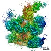
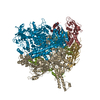
A: Minichromosome maintenance protein MCM, Cell division control protein 21 B: Minichromosome maintenance protein MCM, Cell division control protein 21 hetero molecules
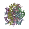
A: Minichromosome maintenance protein MCM, Cell division control protein 21 B: Minichromosome maintenance protein MCM, Cell division control protein 21 hetero molecules
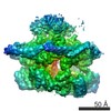
A: Minichromosome maintenance protein MCM, Cell division control protein 21 B: Minichromosome maintenance protein MCM, Cell division control protein 21 hetero molecules
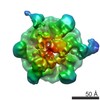
A: Minichromosome maintenance protein MCM, Cell division control protein 21 B: Minichromosome maintenance protein MCM, Cell division control protein 21 hetero molecules
In the structure databanks used in Yorodumi, some data are registered as the other names, "COVID-19 virus" and "2019-nCoV". Here are the details of the virus and the list of structure data.
Jan 31, 2019. EMDB accession codes are about to change! (news from PDBe EMDB page)
EMDB accession codes are about to change! (news from PDBe EMDB page)
The allocation of 4 digits for EMDB accession codes will soon come to an end. Whilst these codes will remain in use, new EMDB accession codes will include an additional digit and will expand incrementally as the available range of codes is exhausted. The current 4-digit format prefixed with “EMD-” (i.e. EMD-XXXX) will advance to a 5-digit format (i.e. EMD-XXXXX), and so on. It is currently estimated that the 4-digit codes will be depleted around Spring 2019, at which point the 5-digit format will come into force.
The EM Navigator/Yorodumi systems omit the EMD- prefix.
Related info.:Q: What is EMD? / ID/Accession-code notation in Yorodumi/EM Navigator
Yorodumi is a browser for structure data from EMDB, PDB, SASBDB, etc.
This page is also the successor to EM Navigator detail page, and also detail information page/front-end page for Omokage search.
The word "yorodu" (or yorozu) is an old Japanese word meaning "ten thousand". "mi" (miru) is to see.
Related info.:EMDB / PDB / SASBDB / Comparison of 3 databanks / Yorodumi Search / Aug 31, 2016. New EM Navigator & Yorodumi / Yorodumi Papers / Jmol/JSmol / Function and homology information / Changes in new EM Navigator and Yorodumi
 Movie
Movie Controller


 Open data
 Basic information
 Components
 Keywords
 Function and homology information

 Authors
 Citation
 Structure visualization
 Downloads & links
 Other downloads


 PDBj
PDBj


 Assembly
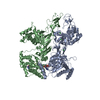


 Mass: 427.201 Da / Num. of mol.: 2 / Source method: obtained synthetically / Formula: C10H15N5O10P2 / Comment: ADP, energy-carrying molecule*YM
 Mass: 24.305 Da / Num. of mol.: 2 / Source method: obtained synthetically / Formula: Mg
 Mass: 65.409 Da / Num. of mol.: 2 / Source method: obtained synthetically / Formula: Zn
 Mass: 35.453 Da / Num. of mol.: 6 / Source method: obtained synthetically / Formula: Cl
 Sample preparation
 / Beamline: 22-ID / Wavelength: 1 Å
 Processing