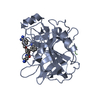
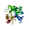
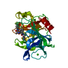
Entry Database : PDB / ID : 4r0iTitle CRYSTAL STRUCTURE of MATRIPTASE in COMPLEX WITH INHIBITOR SERINE PROTEASE, MATRIPTASE, MEMBRANE-TYPE SERINE PROTEASE 1, MT-SP1 Suppressor of tumorigenicity 14 protein Keywords / / / / / Function / homology Function Domain/homology Component
/ / / / / / / / / / / / / / / / / / / / / / / / / / / / / / / / / / / / / / / / / / / / / / / / / / / / / / / / / / / / / Biological species Homo sapiens (human)Method / / Resolution : 1.9 Å Authors Rao, K.N. / Ashok, K.N. / Chakshusmathi, G. / Rajeev, G. / Subramanya, H. Journal : Bioorg.Med.Chem.Lett. / Year : 2015Title : Discovery of O-(3-carbamimidoylphenyl)-l-serine amides as matriptase inhibitors using a fragment-linking approachAuthors : Goswami, R. / Wohlfahrt, G. / Mukherjee, S. / Ghadiyaram, C. / Nagaraj, J. / Satyam, L.K. / Subbarao, K. / Gopinath, S. / Krishnamurthy, N.R. / Subramanya, H.S. / Ramachandra, M. History Deposition Jul 31, 2014 Deposition site / Processing site Revision 1.0 Feb 11, 2015 Provider / Type Revision 1.1 Nov 8, 2023 Group Data collection / Database references ... Data collection / Database references / Derived calculations / Refinement description Category chem_comp_atom / chem_comp_bond ... chem_comp_atom / chem_comp_bond / database_2 / pdbx_initial_refinement_model / struct_site Item _database_2.pdbx_DOI / _database_2.pdbx_database_accession ... _database_2.pdbx_DOI / _database_2.pdbx_database_accession / _struct_site.pdbx_auth_asym_id / _struct_site.pdbx_auth_comp_id / _struct_site.pdbx_auth_seq_id
Show all Show less
 Movie
Movie Controller


 Open data
 Basic information
 Components
 Keywords
 HYDROLASE /
  Function and homology information
 Authors
 Citation
 Structure visualization
 Downloads & links
 Other downloads
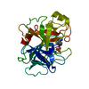


 PDBj
PDBj
 Assembly


 Mass: 523.647 Da / Num. of mol.: 1 / Source method: obtained synthetically / Formula: C27H33N5O4S
 Mass: 18.015 Da / Num. of mol.: 83 / Source method: isolated from a natural source / Formula: H2O
 Sample preparation
 Processing