 Movie
Movie Controller

[English] 日本語
 Yorodumi
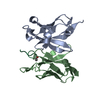
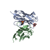
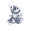
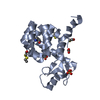
- PDB-4qyo: Crystal Structure of anti-MSP2 Fv fragment (mAb6D8)in complex wit... -
+ Open data

- Basic information
| Entry | Database: PDB / ID: 4qyo | ||||||
|---|---|---|---|---|---|---|---|
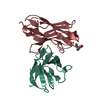
| Title | Crystal Structure of anti-MSP2 Fv fragment (mAb6D8)in complex with MSP2 14-22 | ||||||
 Components Components |
| ||||||
 Keywords Keywords |  IMMUNE SYSTEM / Immunoglobulin fold / N-TERMINAL MSP2 / unstructured antigen IMMUNE SYSTEM / Immunoglobulin fold / N-TERMINAL MSP2 / unstructured antigen | ||||||
| Function / homology |  Function and homology information Function and homology information | ||||||
| Biological species |  Mus musculus (house mouse) Mus musculus (house mouse) Plasmodium falciparum K1 (eukaryote) Plasmodium falciparum K1 (eukaryote) | ||||||
| Method | X-RAY DIFFRACTION / SYNCHROTRON / MOLECULAR REPLACEMENT / Resolution: 1.208 Å | ||||||
 Authors Authors | Morales, R.A.V. / MacRaild, C.A. / Seow, J. / Bankala, K. / Drinkwater, N. / McGowan, S. / Rouet, R. / Christ, D. / Anders, R.F. / Norton, R.S. | ||||||
 Citation Citation | Journal: Sci Rep / Year: 2015 Title: Structural basis for epitope masking and strain specificity of a conserved epitope in an intrinsically disordered malaria vaccine candidate. Authors: Morales, R.A. / MacRaild, C.A. / Seow, J. / Krishnarjuna, B. / Drinkwater, N. / Rouet, R. / Anders, R.F. / Christ, D. / McGowan, S. / Norton, R.S. | ||||||
| History |
|
- Structure visualization
| Structure viewer | Molecule: MolmilJmol/JSmol |
|---|
- Downloads & links
-Download
| PDBx/mmCIF format | 4qyo.cif.gz | 155.2 KB | Display | PDBx/mmCIF format |
|---|---|---|---|---|
| PDB format | pdb4qyo.ent.gz | 125.2 KB | Display | PDB format |
| PDBx/mmJSON format | 4qyo.json.gz | Tree view | PDBx/mmJSON format | |
| Others |  Other downloads Other downloads |
-Validation report
| Arichive directory | https://data.pdbj.org/pub/pdb/validation_reports/qy/4qyoftp://data.pdbj.org/pub/pdb/validation_reports/qy/4qyo | HTTPS FTP |
|---|
-Related structure data
| Related structure data |  4qxtC  4qy8C  4r3sC 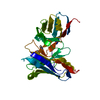 3gm0S C: citing same article ( S: Starting model for refinement |
|---|---|
| Similar structure data |


-Links
 PDBj
PDBj

- Assembly
| Deposited unit | 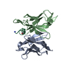
| ||||||||
|---|---|---|---|---|---|---|---|---|---|
| 1 |
| ||||||||
| Unit cell |
|
-Components
| #1: Antibody | Mass: 12489.899 Da / Num. of mol.: 1 Source method: isolated from a genetically manipulated source Source: (gene. exp.) Mus musculus (house mouse) / Production host:  Escherichia coli (E. coli) Escherichia coli (E. coli) |
|---|---|
| #2: Antibody | Mass: 11959.071 Da / Num. of mol.: 1 Source method: isolated from a genetically manipulated source Source: (gene. exp.) Mus musculus (house mouse) / Production host: Escherichia coli (E. coli) |
| #3: Protein/peptide | Mass: 1150.337 Da / Num. of mol.: 1 / Fragment: UNP RESIDUES 33-41 / Source method: obtained synthetically / Source: (synth.) Plasmodium falciparum K1 (eukaryote) / References: UniProt: Q03643 |
| #4: Water | ChemComp-HOH / Water Mass: 18.015 Da / Num. of mol.: 377 / Source method: isolated from a natural source / Formula: H2O Mass: 18.015 Da / Num. of mol.: 377 / Source method: isolated from a natural source / Formula: H2O |
-Experimental details
-Experiment
| Experiment | Method: X-RAY DIFFRACTION / Number of used crystals: 1 |
|---|
- Sample preparation
| Crystal | Density Matthews: 2.08 Å3/Da / Density % sol: 40.92 % |
|---|---|
| Crystal grow | Temperature: 293 K / Method: vapor diffusion, hanging drop / pH: 4.7 Details: 22% PEG 8000, 0.2M NaCl, 0.1M NaOAc, pH 4.7, VAPOR DIFFUSION, HANGING DROP, temperature 293K |
-Data collection
| Diffraction | Mean temperature: 100 K |
|---|---|
| Diffraction source | Source: SYNCHROTRON / Site: Australian Synchrotron  / Beamline: MX1 / Wavelength: 0.9537 Å / Beamline: MX1 / Wavelength: 0.9537 Å |
| Detector | Type: ADSC QUANTUM 210r / Detector: CCD / Date: Mar 29, 2014 |
| Radiation | Protocol: SINGLE WAVELENGTH / Monochromatic (M) / Laue (L): M / Scattering type: x-ray |
| Radiation wavelength | Wavelength: 0.9537 Å / Relative weight: 1 |
| Reflection | Resolution: 1.208→41.864 Å / Num. all: 62158 / Num. obs: 62158 / % possible obs: 96.75 % |
| Reflection shell | Resolution: 1.208→1.251 Å / Mean I/σ(I) obs: 1.5 / Num. unique all: 5342 / % possible all: 83.25 |
- Processing
| Software |
| ||||||||||||||||||||||||||||||||||||||||||||||||||||||||||||||||||||||||||||||||||||||||||||||||||||||||||||||||||||||||||||||||||||||||||
|---|---|---|---|---|---|---|---|---|---|---|---|---|---|---|---|---|---|---|---|---|---|---|---|---|---|---|---|---|---|---|---|---|---|---|---|---|---|---|---|---|---|---|---|---|---|---|---|---|---|---|---|---|---|---|---|---|---|---|---|---|---|---|---|---|---|---|---|---|---|---|---|---|---|---|---|---|---|---|---|---|---|---|---|---|---|---|---|---|---|---|---|---|---|---|---|---|---|---|---|---|---|---|---|---|---|---|---|---|---|---|---|---|---|---|---|---|---|---|---|---|---|---|---|---|---|---|---|---|---|---|---|---|---|---|---|---|---|---|---|
| Refinement | Method to determine structure: MOLECULAR REPLACEMENT Starting model: PDB ID 3GM0 Resolution: 1.208→41.864 Å / SU ML: 0.11 / σ(F): 1.34 / Phase error: 17.6 / Stereochemistry target values: Engh & Huber
| ||||||||||||||||||||||||||||||||||||||||||||||||||||||||||||||||||||||||||||||||||||||||||||||||||||||||||||||||||||||||||||||||||||||||||
| Solvent computation | Shrinkage radii: 0.9 Å / VDW probe radii: 1.11 Å / Solvent model: FLAT BULK SOLVENT MODEL | ||||||||||||||||||||||||||||||||||||||||||||||||||||||||||||||||||||||||||||||||||||||||||||||||||||||||||||||||||||||||||||||||||||||||||
| Refinement step | Cycle: LAST / Resolution: 1.208→41.864 Å
| ||||||||||||||||||||||||||||||||||||||||||||||||||||||||||||||||||||||||||||||||||||||||||||||||||||||||||||||||||||||||||||||||||||||||||
| Refine LS restraints |
| ||||||||||||||||||||||||||||||||||||||||||||||||||||||||||||||||||||||||||||||||||||||||||||||||||||||||||||||||||||||||||||||||||||||||||
| LS refinement shell | Refine-ID: X-RAY DIFFRACTION / Total num. of bins used: 22
| ||||||||||||||||||||||||||||||||||||||||||||||||||||||||||||||||||||||||||||||||||||||||||||||||||||||||||||||||||||||||||||||||||||||||||
| Refinement TLS params. | Method: refined / Origin x: 19.8775 Å / Origin y: 17.242 Å / Origin z: 33.177 Å
| ||||||||||||||||||||||||||||||||||||||||||||||||||||||||||||||||||||||||||||||||||||||||||||||||||||||||||||||||||||||||||||||||||||||||||
| Refinement TLS group | Selection details: ALL |
