 Movie
Movie Controller


+ Open data

- Basic information
| Entry | Database: PDB / ID: 3gm0 | ||||||
|---|---|---|---|---|---|---|---|
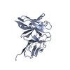
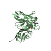
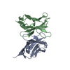
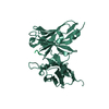
| Title | Anti-methamphetamine single chain Fv in complex with MDMA | ||||||
 Components Components | anti-methamphetamine single chain Fv | ||||||
 Keywords Keywords |  IMMUNE SYSTEM / anti-methamphetamine antibody / single chain Fv / Therapeutic antibody IMMUNE SYSTEM / anti-methamphetamine antibody / single chain Fv / Therapeutic antibody | ||||||
| Function / homology | Immunoglobulins / Immunoglobulin-like / Sandwich / Mainly Beta / Chem-B41 Function and homology information Function and homology information | ||||||
| Biological species |  Mus musculus (house mouse) Mus musculus (house mouse) | ||||||
| Method | X-RAY DIFFRACTION / SYNCHROTRON / Resolution: 2.4 Å | ||||||
 Authors Authors | Celikel, R. / Peterson, E.C. / Owens, M. / Varughese, K.I. | ||||||
 Citation Citation | Journal: Protein Sci. / Year: 2009 Title: Crystal structures of a therapeutic single chain antibody in complex with two drugs of abuse-Methamphetamine and 3,4-methylenedioxymethamphetamine. Authors: Celikel, R. / Peterson, E.C. / Owens, S.M. / Varughese, K.I. | ||||||
| History |
|
- Structure visualization
| Structure viewer | Molecule: MolmilJmol/JSmol |
|---|
- Downloads & links
-Download
| PDBx/mmCIF format | 3gm0.cif.gz | 56.3 KB | Display | PDBx/mmCIF format |
|---|---|---|---|---|
| PDB format | pdb3gm0.ent.gz | 43.5 KB | Display | PDB format |
| PDBx/mmJSON format | 3gm0.json.gz | Tree view | PDBx/mmJSON format | |
| Others |  Other downloads Other downloads |
-Validation report
| Arichive directory | https://data.pdbj.org/pub/pdb/validation_reports/gm/3gm0ftp://data.pdbj.org/pub/pdb/validation_reports/gm/3gm0 | HTTPS FTP |
|---|
-Related structure data


-Links
 PDBj
PDBj

- Assembly
| Deposited unit | 
| ||||||||
|---|---|---|---|---|---|---|---|---|---|
| 1 |
| ||||||||
| Unit cell |
|
-Components
| #1: Antibody | Mass: 27789.309 Da / Num. of mol.: 1 Source method: isolated from a genetically manipulated source Source: (gene. exp.) Mus musculus (house mouse) / Gene: IgG / Plasmid: pPICZ-alpha / Production host:  Pichia pastoris (fungus) / Strain (production host): KM71H Pichia pastoris (fungus) / Strain (production host): KM71H |
|---|---|
| #2: Chemical | ChemComp-B41 / (MDMA  Mass: 193.242 Da / Num. of mol.: 1 / Source method: obtained synthetically / Formula: C11H15NO2 Mass: 193.242 Da / Num. of mol.: 1 / Source method: obtained synthetically / Formula: C11H15NO2 |
| #3: Water | ChemComp-HOH / Water Mass: 18.015 Da / Num. of mol.: 77 / Source method: isolated from a natural source / Formula: H2O Mass: 18.015 Da / Num. of mol.: 77 / Source method: isolated from a natural source / Formula: H2O |
| Sequence details | THE PROTEIN WAS CRYSTALLIZED AS A FUSION PROTEIN WHERE TWO CHAINS, CHAIN H AND CHAIN L, WERE FUSED ...THE PROTEIN WAS CRYSTALLIZ |
-Experimental details
-Experiment
| Experiment | Method: X-RAY DIFFRACTION / Number of used crystals: 1 |
|---|
- Sample preparation
| Crystal | Density Matthews: 1.93 Å3/Da / Density % sol: 36.35 % |
|---|---|
| Crystal grow | Temperature: 287 K / Method: vapor diffusion / pH: 8.3 Details: 1.15M sodium Citrate, 0.28M Imidazole malate, pH 8.3, VAPOR DIFFUSION, temperature 287K |
-Data collection
| Diffraction | Mean temperature: 100 K |
|---|---|
| Diffraction source | Source: SYNCHROTRON / Site: SSRL  / Beamline: BL9-1 / Wavelength: 0.97945 Å / Beamline: BL9-1 / Wavelength: 0.97945 Å |
| Detector | Type: ADSC QUANTUM 315 / Detector: CCD / Date: Jun 18, 2008 / Details: single crystal bent monochromator |
| Radiation | Monochromator: single crystal bent monochromator / Protocol: SINGLE WAVELENGTH / Monochromatic (M) / Laue (L): M / Scattering type: x-ray |
| Radiation wavelength | Wavelength: 0.97945 Å / Relative weight: 1 |
| Reflection | Resolution: 2.4→32.4 Å / Num. all: 8316 / Num. obs: 8316 / % possible obs: 99.8 % / Observed criterion σ(F): 0 / Redundancy: 3.75 % / Rsym value: 0.045 |
| Reflection shell | Resolution: 2.4→2.58 Å / Redundancy: 3.7 % / Rsym value: 0.21 / % possible all: 99.5 |
- Processing
| Software |
| ||||||||||||||||||||||||||||||||||||
|---|---|---|---|---|---|---|---|---|---|---|---|---|---|---|---|---|---|---|---|---|---|---|---|---|---|---|---|---|---|---|---|---|---|---|---|---|---|
| Refinement | Resolution: 2.4→15 Å / Occupancy max: 1 / Occupancy min: 1 / σ(F): 0
| ||||||||||||||||||||||||||||||||||||
| Solvent computation | Bsol: 66.944 Å2 | ||||||||||||||||||||||||||||||||||||
| Displacement parameters | Biso max: 82.68 Å2 / Biso mean: 39.042 Å2 / Biso min: 16.72 Å2
| ||||||||||||||||||||||||||||||||||||
| Refine analyze | Luzzati coordinate error obs: 0.32 Å / Luzzati d res low obs: 5 Å / Luzzati sigma a obs: 0.32 Å | ||||||||||||||||||||||||||||||||||||
| Refinement step | Cycle: LAST / Resolution: 2.4→15 Å
| ||||||||||||||||||||||||||||||||||||
| Refine LS restraints |
| ||||||||||||||||||||||||||||||||||||
| LS refinement shell |
| ||||||||||||||||||||||||||||||||||||
| Xplor file |
|
