acetyltransferase activator activity / regulation of developmental process / MOZ/MORF histone acetyltransferase complex / regulation of hemopoiesis / histone acetyltransferase complex / negative regulation of megakaryocyte differentiation / protein localization to CENP-A containing chromatin / Replacement of protamines by nucleosomes in the male pronucleus / CENP-A containing nucleosome / Packaging Of Telomere Ends ...acetyltransferase activator activity / regulation of developmental process / MOZ/MORF histone acetyltransferase complex / regulation of hemopoiesis / histone acetyltransferase complex / negative regulation of megakaryocyte differentiation / protein localization to CENP-A containing chromatin / Replacement of protamines by nucleosomes in the male pronucleus / CENP-A containing nucleosome / Packaging Of Telomere Ends / Recognition and association of DNA glycosylase with site containing an affected purine / Cleavage of the damaged purine / Deposition of new CENPA-containing nucleosomes at the centromere / Regulation of TP53 Activity through Acetylation / Recognition and association of DNA glycosylase with site containing an affected pyrimidine / Cleavage of the damaged pyrimidine / Inhibition of DNA recombination at telomere / Meiotic synapsis / telomere organization / RNA Polymerase I Promoter Opening / Assembly of the ORC complex at the origin of replication / SUMOylation of chromatin organization proteins / DNA methylation / Condensation of Prophase Chromosomes / ERCC6 (CSB) and EHMT2 (G9a) positively regulate rRNA expression / SIRT1 negatively regulates rRNA expression / Chromatin modifications during the maternal to zygotic transition (MZT) / HCMV Late Events / PRC2 methylates histones and DNA / Defective pyroptosis / HDACs deacetylate histones / RNA Polymerase I Promoter Escape / Nonhomologous End-Joining (NHEJ) / Transcriptional regulation by small RNAs / Formation of the beta-catenin:TCF transactivating complex / RUNX1 regulates genes involved in megakaryocyte differentiation and platelet function / Activated PKN1 stimulates transcription of AR (androgen receptor) regulated genes KLK2 and KLK3 / NoRC negatively regulates rRNA expression / G2/M DNA damage checkpoint / B-WICH complex positively regulates rRNA expression / HDMs demethylate histones / DNA Damage/Telomere Stress Induced Senescence / PKMTs methylate histone lysines / RMTs methylate histone arginines / Meiotic recombination / Pre-NOTCH Transcription and Translation / Activation of anterior HOX genes in hindbrain development during early embryogenesis / HCMV Early Events / Transcriptional regulation of granulopoiesis / structural constituent of chromatin / nucleosome / nucleosome assembly / Recruitment and ATM-mediated phosphorylation of repair and signaling proteins at DNA double strand breaks / RUNX1 regulates transcription of genes involved in differentiation of HSCs / chromatin organization / HATs acetylate histones / Processing of DNA double-strand break ends / Senescence-Associated Secretory Phenotype (SASP) / Oxidative Stress Induced Senescence / Estrogen-dependent gene expression / chromosome, telomeric region / chromatin remodeling / protein heterodimerization activity / Amyloid fiber formation / regulation of DNA-templated transcription / regulation of transcription by RNA polymerase II / positive regulation of DNA-templated transcription / protein-containing complex / DNA binding / RNA binding / extracellular exosome / extracellular region / nucleoplasm / membrane / metal ion binding / nucleus / plasma membrane / cytosol / cytoplasm Similarity search - Function
Resolution: 1.94→34.43 Å / SU ML: 0.12 / σ(F): 1.37 / Phase error: 18.93 / Stereochemistry target values: MAXIMUM LIKELIHOOD / Details: HYDROGENS HAVE BEEN ADDED IN THE RIDING POSITIONS
Rfactor
Num. reflection
% reflection
Rfree
0.213
966
5.14 %
Rwork
0.175
-
-
obs
0.177
18780
99.7 %
all
-
18780
-
Solvent computation
Shrinkage radii: 0.9 Å / VDW probe radii: 1.11 Å / Solvent model: FLAT BULK SOLVENT MODEL
Refinement step
Cycle: LAST / Resolution: 1.94→34.43 Å
Protein
Nucleic acid
Ligand
Solvent
Total
Num. atoms
995
0
0
143
1138
Refine LS restraints
Refine-ID
Type
Dev ideal
Number
X-RAY DIFFRACTION
f_bond_d
0.007
1014
X-RAY DIFFRACTION
f_angle_d
0.95
1361
X-RAY DIFFRACTION
f_dihedral_angle_d
12.875
392
X-RAY DIFFRACTION
f_chiral_restr
0.066
146
X-RAY DIFFRACTION
f_plane_restr
0.004
179
LS refinement shell
Resolution (Å)
Rfactor Rfree
Num. reflection Rfree
Rfactor Rwork
Num. reflection Rwork
Refine-ID
% reflection obs (%)
1.94-2.0435
0.2092
129
0.1709
2475
X-RAY DIFFRACTION
98
2.0435-2.1715
0.2178
124
0.1631
2494
X-RAY DIFFRACTION
100
2.1715-2.3392
0.1827
152
0.17
2496
X-RAY DIFFRACTION
100
2.3392-2.5745
0.1862
142
0.1724
2509
X-RAY DIFFRACTION
100
2.5745-2.9469
0.2323
136
0.1879
2554
X-RAY DIFFRACTION
100
2.9469-3.712
0.2174
128
0.1751
2584
X-RAY DIFFRACTION
100
3.712-34.4334
0.2214
155
0.1763
2702
X-RAY DIFFRACTION
100
Refinement TLS params.
Method: refined / Refine-ID: X-RAY DIFFRACTION
ID
L11 (°2)
L12 (°2)
L13 (°2)
L22 (°2)
L23 (°2)
L33 (°2)
S11 (Å °)
S12 (Å °)
S13 (Å °)
S21 (Å °)
S22 (Å °)
S23 (Å °)
S31 (Å °)
S32 (Å °)
S33 (Å °)
T11 (Å2)
T12 (Å2)
T13 (Å2)
T22 (Å2)
T23 (Å2)
T33 (Å2)
Origin x (Å)
Origin y (Å)
Origin z (Å)
1
5.4108
-1.244
0.0155
6.0042
0.5356
4.3278
-0.1516
-2.1388
-0.0482
1.3247
-0.03
-1.0712
0.6523
1.1199
-0.0107
0.2211
0.1663
-0.111
0.8282
0.176
0.2771
25.4797
84.2492
100.5969
2
5.8942
-1.547
-1.8234
3.8617
0.2883
2.0082
-0.1071
-0.7629
-0.2557
0.2265
0.0497
0.3069
-0.0514
0.0281
0.0526
0.1793
0.0099
0.0498
0.278
-0.0039
0.1789
4.0124
90.7519
99.5984
3
3.5762
-0.5241
-1.2526
1.9787
0.5419
2.5334
-0.2054
-0.6827
-0.7573
0.2374
0.192
0.2145
0.357
0.2452
-0.0042
0.2128
0.0298
0.0417
0.2858
0.0893
0.2476
14.5492
84.0801
96.1942
4
7.2357
-1.752
-1.4959
2.913
0.7923
2.363
0.3091
-0.3135
1.0751
0.0648
0.0151
-0.4337
-0.259
0.1843
-0.2866
0.1648
-0.0345
0.0235
0.1795
-0.0161
0.2065
17.7847
96.1331
92.6837
5
5.0365
4.4141
5.2693
4.4845
3.9937
6.1669
-0.087
-0.769
-1.6659
-0.3926
0.9089
-0.6738
1.1338
0.8484
-0.6526
0.5834
0.1729
-0.0941
0.8354
0.0089
1.0018
31.2406
77.4544
89.4081
6
6.8979
-2.2711
-0.0967
6.5191
-4.9717
4.3363
0.072
0.7045
0.5537
-0.501
-0.1674
-0.2522
0.3496
-0.677
0.1083
0.2076
0.0513
0.0461
0.3938
-0.0227
0.1733
-1.7363
98.0137
94.6083
Refinement TLS group
ID
Refine-ID
Refine TLS-ID
Selection details
1
X-RAY DIFFRACTION
1
chain 'A' and (resid4through22 )
2
X-RAY DIFFRACTION
2
chain 'A' and (resid23through49 )
3
X-RAY DIFFRACTION
3
chain 'A' and (resid50through82 )
4
X-RAY DIFFRACTION
4
chain 'A' and (resid83through112 )
5
X-RAY DIFFRACTION
5
chain 'A' and (resid113through117 )
6
X-RAY DIFFRACTION
6
chain 'B' and (resid6through13 )
+
About Yorodumi
-
News
-
Feb 9, 2022. New format data for meta-information of EMDB entries
New format data for meta-information of EMDB entries
Version 3 of the EMDB header file is now the official format.
The previous official version 1.9 will be removed from the archive.
In the structure databanks used in Yorodumi, some data are registered as the other names, "COVID-19 virus" and "2019-nCoV". Here are the details of the virus and the list of structure data.
Jan 31, 2019. EMDB accession codes are about to change! (news from PDBe EMDB page)
EMDB accession codes are about to change! (news from PDBe EMDB page)
The allocation of 4 digits for EMDB accession codes will soon come to an end. Whilst these codes will remain in use, new EMDB accession codes will include an additional digit and will expand incrementally as the available range of codes is exhausted. The current 4-digit format prefixed with “EMD-” (i.e. EMD-XXXX) will advance to a 5-digit format (i.e. EMD-XXXXX), and so on. It is currently estimated that the 4-digit codes will be depleted around Spring 2019, at which point the 5-digit format will come into force.
The EM Navigator/Yorodumi systems omit the EMD- prefix.
Related info.:Q: What is EMD? / ID/Accession-code notation in Yorodumi/EM Navigator
Yorodumi is a browser for structure data from EMDB, PDB, SASBDB, etc.
This page is also the successor to EM Navigator detail page, and also detail information page/front-end page for Omokage search.
The word "yorodu" (or yorozu) is an old Japanese word meaning "ten thousand". "mi" (miru) is to see.
Related info.:EMDB / PDB / SASBDB / Comparison of 3 databanks / Yorodumi Search / Aug 31, 2016. New EM Navigator & Yorodumi / Yorodumi Papers / Jmol/JSmol / Function and homology information / Changes in new EM Navigator and Yorodumi
 Movie
Movie Controller

 Yorodumi
 Open data
 Basic information
 Components
 Keywords
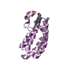
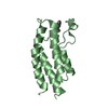
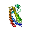
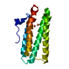
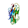
 PROTEIN BINDING / bromodomain-PHD finger protein 1 (BRPF1) / histone acetyltransferase (HAT) / monocytic leukemia zinc-finger (MOZ) /
  Function and homology information
 Authors
 Citation
 Structure visualization
 Downloads & links
 Other downloads


 PDBj
PDBj


 Assembly

 Mass: 18.015 Da / Num. of mol.: 143 / Source method: isolated from a natural source / Formula: H2O
 Sample preparation
 / Beamline: X29A / Wavelength: 1.075
  Processing