negative regulation of cellular response to oxidative stress / TCF dependent signaling in response to WNT / Degradation of AXIN / positive regulation of protein polyubiquitination / Regulation of PTEN stability and activity / Conversion from APC/C:Cdc20 to APC/C:Cdh1 in late anaphase / poly-ADP-D-ribose binding / Inactivation of APC/C via direct inhibition of the APC/C complex / APC/C:Cdc20 mediated degradation of mitotic proteins / Aberrant regulation of mitotic exit in cancer due to RB1 defects ...negative regulation of cellular response to oxidative stress / TCF dependent signaling in response to WNT / Degradation of AXIN / positive regulation of protein polyubiquitination / Regulation of PTEN stability and activity / Conversion from APC/C:Cdc20 to APC/C:Cdh1 in late anaphase / poly-ADP-D-ribose binding / Inactivation of APC/C via direct inhibition of the APC/C complex / APC/C:Cdc20 mediated degradation of mitotic proteins / Aberrant regulation of mitotic exit in cancer due to RB1 defects / (E3-independent) E2 ubiquitin-conjugating enzyme / Phosphorylation of the APC/C / Signaling by BMP / Ub-specific processing proteases / E2 ubiquitin-conjugating enzyme / ubiquitin conjugating enzyme activity / Regulation of APC/C activators between G1/S and early anaphase / negative regulation of BMP signaling pathway / Transcriptional Regulation by VENTX / protein K48-linked ubiquitination / protein autoubiquitination / ubiquitin ligase complex / negative regulation of TORC1 signaling / APC/C:Cdc20 mediated degradation of Cyclin B / APC-Cdc20 mediated degradation of Nek2A / Synthesis of active ubiquitin: roles of E1 and E2 enzymes / TICAM1, RIP1-mediated IKK complex recruitment / IKK complex recruitment mediated by RIP1 / Autodegradation of Cdh1 by Cdh1:APC/C / APC/C:Cdc20 mediated degradation of Securin / positive regulation of protein ubiquitination / Assembly of the pre-replicative complex / Negative regulators of DDX58/IFIH1 signaling / Cdc20:Phospho-APC/C mediated degradation of Cyclin A / Peroxisomal protein import / Downregulation of SMAD2/3:SMAD4 transcriptional activity / Regulation of TNFR1 signaling / APC/C:Cdh1 mediated degradation of Cdc20 and other APC/C:Cdh1 targeted proteins in late mitosis/early G1 / RING-type E3 ubiquitin transferase / Oxygen-dependent proline hydroxylation of Hypoxia-inducible Factor Alpha / Inactivation of CSF3 (G-CSF) signaling / CDK-mediated phosphorylation and removal of Cdc6 / cellular response to hydrogen peroxide / CLEC7A (Dectin-1) signaling / Wnt signaling pathway / FCERI mediated NF-kB activation / protein polyubiquitination / ubiquitin-protein transferase activity / Separation of Sister Chromatids / positive regulation of canonical Wnt signaling pathway / ubiquitin protein ligase activity / Ovarian tumor domain proteases / Antigen processing: Ubiquitination & Proteasome degradation / Downstream TCR signaling / E3 ubiquitin ligases ubiquitinate target proteins / Neddylation / ubiquitin-dependent protein catabolic process / Senescence-Associated Secretory Phenotype (SASP) / proteasome-mediated ubiquitin-dependent protein catabolic process / ubiquitin protein ligase binding / negative regulation of transcription by RNA polymerase II / enzyme binding / protein-containing complex / zinc ion binding / nucleoplasm / ATP binding / nucleus / plasma membrane / cytosol / cytoplasm Similarity search - Function
E3 ubiquitin-protein ligase RNF146 / RNF146, RING finger, HC subclass / Signal recognition particle alu RNA binding heterodimer, srp9/1 - #50 / WWE domain, subgroup / Domain in Deltex and TRIP12 homologues. Possibly involved in regulation of ubiquitin-mediated proteolysis. / Signal recognition particle alu RNA binding heterodimer, srp9/1 / WWE domain / WWE domain superfamily / WWE domain / WWE domain profile. ...E3 ubiquitin-protein ligase RNF146 / RNF146, RING finger, HC subclass / Signal recognition particle alu RNA binding heterodimer, srp9/1 - #50 / WWE domain, subgroup / Domain in Deltex and TRIP12 homologues. Possibly involved in regulation of ubiquitin-mediated proteolysis. / Signal recognition particle alu RNA binding heterodimer, srp9/1 / WWE domain / WWE domain superfamily / WWE domain / WWE domain profile. / Ubiquitin Conjugating Enzyme / Ubiquitin Conjugating Enzyme / Zinc finger, C3HC4 type (RING finger) / Ubiquitin-conjugating enzyme, active site / Ubiquitin-conjugating (UBC) active site signature. / Ubiquitin-conjugating enzyme E2, catalytic domain homologues / Ubiquitin-conjugating enzyme E2 / Ubiquitin-conjugating enzyme / Ubiquitin-conjugating (UBC) core domain profile. / Ubiquitin-conjugating enzyme/RWD-like / Zinc finger, RING-type, conserved site / Zinc finger RING-type signature. / Ring finger / Zinc finger RING-type profile. / Zinc finger, RING-type / Zinc finger, RING/FYVE/PHD-type / Roll / 2-Layer Sandwich / Alpha Beta Similarity search - Domain/homology
Mass: 18.015 Da / Num. of mol.: 371 / Source method: isolated from a natural source / Formula: H2O
-
Experimental details
-
Experiment
Experiment
Method: X-RAY DIFFRACTION / Number of used crystals: 1
-
Sample preparation
Crystal
Density Matthews: 2.75 Å3/Da / Density % sol: 55.25 %
Crystal grow
Temperature: 295 K / Method: vapor diffusion, hanging drop / pH: 7 Details: 0.8 M sodium citrate, 80 mM Tris HCl pH7.0, 160 mM NaCl, 4 mMDTT, 20 mM trimethylamin HCl, VAPOR DIFFUSION, HANGING DROP, temperature 295K
-
Data collection
Diffraction
Mean temperature: 100 K
Diffraction source
Source: SYNCHROTRON / Site: ALS / Beamline: 8.2.1 / Wavelength: 1.283 Å
Resolution: 1.9→1.96 Å / Χ2: 0.807 / % possible all: 71.4
-
Processing
Software
Name
Version
Classification
NB
DENZO
datareduction
SCALEPACK
datascaling
REFMAC
5.8.0049
refinement
PDB_EXTRACT
3.14
dataextraction
HKL-2000
datacollection
HKL-2000
datareduction
SOLVE
phasing
Refinement
Method to determine structure: SAD / Resolution: 1.9→50 Å / Cor.coef. Fo:Fc: 0.964 / Cor.coef. Fo:Fc free: 0.948 / SU B: 4.656 / SU ML: 0.08 / Cross valid method: THROUGHOUT / σ(F): 0 / ESU R: 0.145 / ESU R Free: 0.135 / Stereochemistry target values: MAXIMUM LIKELIHOOD Details: HYDROGENS HAVE BEEN ADDED IN THE RIDING POSITIONS U VALUES : WITH TLS ADDED
Rfactor
Num. reflection
% reflection
Selection details
Rfree
0.222
2994
5.1 %
RANDOM
Rwork
0.1864
-
-
-
all
0.1882
58752
-
-
obs
0.1882
55779
94.94 %
-
Solvent computation
Ion probe radii: 0.8 Å / Shrinkage radii: 0.8 Å / VDW probe radii: 1.2 Å / Solvent model: MASK
In the structure databanks used in Yorodumi, some data are registered as the other names, "COVID-19 virus" and "2019-nCoV". Here are the details of the virus and the list of structure data.
Jan 31, 2019. EMDB accession codes are about to change! (news from PDBe EMDB page)
EMDB accession codes are about to change! (news from PDBe EMDB page)
The allocation of 4 digits for EMDB accession codes will soon come to an end. Whilst these codes will remain in use, new EMDB accession codes will include an additional digit and will expand incrementally as the available range of codes is exhausted. The current 4-digit format prefixed with “EMD-” (i.e. EMD-XXXX) will advance to a 5-digit format (i.e. EMD-XXXXX), and so on. It is currently estimated that the 4-digit codes will be depleted around Spring 2019, at which point the 5-digit format will come into force.
The EM Navigator/Yorodumi systems omit the EMD- prefix.
Related info.:Q: What is EMD? / ID/Accession-code notation in Yorodumi/EM Navigator
Yorodumi is a browser for structure data from EMDB, PDB, SASBDB, etc.
This page is also the successor to EM Navigator detail page, and also detail information page/front-end page for Omokage search.
The word "yorodu" (or yorozu) is an old Japanese word meaning "ten thousand". "mi" (miru) is to see.
Related info.:EMDB / PDB / SASBDB / Comparison of 3 databanks / Yorodumi Search / Aug 31, 2016. New EM Navigator & Yorodumi / Yorodumi Papers / Jmol/JSmol / Function and homology information / Changes in new EM Navigator and Yorodumi
 Movie
Movie Controller


 Open data
 Basic information
 Components
 Keywords
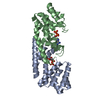
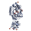
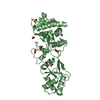
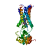
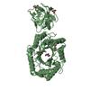
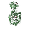
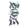
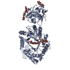
 LIGASE / Protein poly(ADP-ribosy)lation /
  Function and homology information

 Authors
 Citation
 Structure visualization
 Downloads & links
 Other downloads
 PDBj
PDBj


 Assembly


 Type: RNA linking / Mass: 559.316 Da / Num. of mol.: 2 / Source method: obtained synthetically / Formula: C15H23N5O14P2
 Mass: 65.409 Da / Num. of mol.: 4 / Source method: obtained synthetically / Formula: Zn
 Mass: 18.015 Da / Num. of mol.: 371 / Source method: isolated from a natural source / Formula: H2O
 Sample preparation
 / Beamline: 8.2.1 / Wavelength: 1.283 Å
 Processing