 Movie
Movie Controller

[English] 日本語
 Yorodumi
- PDB-4qf2: Crystal structure of human BAZ2A PHD zinc finger in the free form -
+ Open data

- Basic information
| Entry | Database: PDB / ID: 4qf2 | ||||||
|---|---|---|---|---|---|---|---|
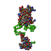
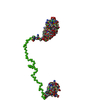
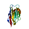
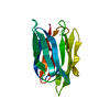
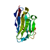
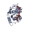
| Title | Crystal structure of human BAZ2A PHD zinc finger in the free form | ||||||
 Components Components | Bromodomain adjacent to zinc finger domain protein 2A | ||||||
 Keywords Keywords |  TRANSCRIPTION / Bromodomain adjacent to zinc finger domain protein 2A / Transcription termination factor I-interacting protein 5 / TTF-I-interacting protein 5 TRANSCRIPTION / Bromodomain adjacent to zinc finger domain protein 2A / Transcription termination factor I-interacting protein 5 / TTF-I-interacting protein 5 | ||||||
| Function / homology |  Function and homology information Function and homology informationNoRC complex / : / : / rDNA heterochromatin / rDNA heterochromatin formation / chromatin silencing complex / RNA polymerase I preinitiation complex assembly / : / negative regulation of transcription by RNA polymerase I / : ...NoRC complex / : / : / rDNA heterochromatin / rDNA heterochromatin formation / chromatin silencing complex / RNA polymerase I preinitiation complex assembly / : / negative regulation of transcription by RNA polymerase I / : / heterochromatin formation / nuclear receptor binding / lysine-acetylated histone binding / NoRC negatively regulates rRNA expression / histone binding / nuclear speck / chromatin remodeling / DNA-templated transcription / nucleolus / regulation of DNA-templated transcription / DNA binding / RNA binding / metal ion binding / nucleus / cytosolSimilarity search - Function | ||||||
| Biological species |  Homo sapiens (human) Homo sapiens (human) | ||||||
| Method | X-RAY DIFFRACTION / SYNCHROTRON / SAD / Resolution: 1.7 Å | ||||||
 Authors Authors | Tallant, C. / Overvoorde, L. / Van Molle, I. / Chirgadze, D.Y. / Ciulli, A. | ||||||
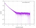
 Citation Citation | Journal: Structure / Year: 2015 Title: Molecular basis of histone tail recognition by human TIP5 PHD finger and bromodomain of the chromatin remodeling complex NoRC. Authors: Cynthia Tallant / Erica Valentini / Oleg Fedorov / Lois Overvoorde / Fleur M Ferguson / Panagis Filippakopoulos / Dmitri I Svergun / Stefan Knapp / Alessio Ciulli /   Abstract: Binding of the chromatin remodeling complex NoRC to RNA complementary to the rDNA promoter mediates transcriptional repression. TIP5, the largest subunit of NoRC, is involved in recruitment to rDNA ...Binding of the chromatin remodeling complex NoRC to RNA complementary to the rDNA promoter mediates transcriptional repression. TIP5, the largest subunit of NoRC, is involved in recruitment to rDNA by interactions with promoter-bound TTF-I, pRNA, and acetylation of H4K16. TIP5 domains that recognize posttranslational modifications on histones are essential for recruitment of NoRC to chromatin, but how these reader modules recognize site-specific histone tails has remained elusive. Here, we report crystal structures of PHD zinc finger and bromodomains from human TIP5 and BAZ2B in free form and bound to H3 and/or H4 histones. PHD finger functions as an independent structural module in recognizing unmodified H3 histone tails, and the bromodomain prefers H3 and H4 acetylation marks followed by a key basic residue, KacXXR. Further low-resolution analyses of PHD-bromodomain modules provide molecular insights into their trans histone tail recognition, required for nucleosome recruitment and transcriptional repression of the NoRC complex. | ||||||
| History |
|
- Structure visualization
| Structure viewer | Molecule: MolmilJmol/JSmol |
|---|
- Downloads & links
-Download
| PDBx/mmCIF format | 4qf2.cif.gz | 97.9 KB | Display | PDBx/mmCIF format |
|---|---|---|---|---|
| PDB format | pdb4qf2.ent.gz | 75.9 KB | Display | PDB format |
| PDBx/mmJSON format | 4qf2.json.gz | Tree view | PDBx/mmJSON format | |
| Others |  Other downloads Other downloads |
-Validation report
| Arichive directory | https://data.pdbj.org/pub/pdb/validation_reports/qf/4qf2ftp://data.pdbj.org/pub/pdb/validation_reports/qf/4qf2 | HTTPS FTP |
|---|
-Related structure data
| Related structure data |  4lz2C 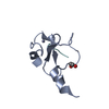 4q6fC  4qbmC 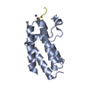 4qc1C 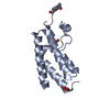 4qc3C  4qf3C C: citing same article ( |
|---|---|
| Similar structure data |


-Links
 PDBj
PDBj


- Assembly
| Deposited unit | 
| ||||||||
|---|---|---|---|---|---|---|---|---|---|
| 1 | 
| ||||||||
| 2 | 
| ||||||||
| Unit cell |
|
-Components
| #1: Protein | Mass: 6597.694 Da / Num. of mol.: 4 / Fragment: unp residues 1673-1728 Source method: isolated from a genetically manipulated source Source: (gene. exp.) Homo sapiens (human) / Gene: BAZ2A, KIAA0314, TIP5 / Plasmid: pET15b / Production host:  Escherichia coli (E. coli) / Strain (production host): BL21(DE3)Rosetta / References: UniProt: Q9UIF9 Escherichia coli (E. coli) / Strain (production host): BL21(DE3)Rosetta / References: UniProt: Q9UIF9#2: Chemical | ChemComp-ZN /   Mass: 65.409 Da / Num. of mol.: 8 / Source method: obtained synthetically / Formula: Zn Mass: 65.409 Da / Num. of mol.: 8 / Source method: obtained synthetically / Formula: Zn#3: Chemical | Phosphate  Mass: 94.971 Da / Num. of mol.: 3 / Source method: obtained synthetically / Formula: PO4 Mass: 94.971 Da / Num. of mol.: 3 / Source method: obtained synthetically / Formula: PO4#4: Chemical | ChemComp-GOL / | Glycerol  Mass: 92.094 Da / Num. of mol.: 1 / Source method: obtained synthetically / Formula: C3H8O3 Mass: 92.094 Da / Num. of mol.: 1 / Source method: obtained synthetically / Formula: C3H8O3#5: Water | ChemComp-HOH / | Water Mass: 18.015 Da / Num. of mol.: 120 / Source method: isolated from a natural source / Formula: H2O Mass: 18.015 Da / Num. of mol.: 120 / Source method: isolated from a natural source / Formula: H2O |
|---|
-Experimental details
-Experiment
| Experiment | Method: X-RAY DIFFRACTION / Number of used crystals: 1 |
|---|
- Sample preparation
| Crystal | Density Matthews: 2.43 Å3/Da / Density % sol: 49.45 % |
|---|---|
| Crystal grow | Temperature: 293 K / Method: vapor diffusion, sitting drop / pH: 8 Details: 2M Na/K phosphate, pH 8, VAPOR DIFFUSION, SITTING DROP, temperature 293K |
-Data collection
| Diffraction | Mean temperature: 100 K | ||||||||||||||||||||||||||||||||||||||||||||||||||||||||||||||||||
|---|---|---|---|---|---|---|---|---|---|---|---|---|---|---|---|---|---|---|---|---|---|---|---|---|---|---|---|---|---|---|---|---|---|---|---|---|---|---|---|---|---|---|---|---|---|---|---|---|---|---|---|---|---|---|---|---|---|---|---|---|---|---|---|---|---|---|---|
| Diffraction source | Source: SYNCHROTRON / Site: Diamond / Beamline: I02 / Wavelength: 1.2822 Å | ||||||||||||||||||||||||||||||||||||||||||||||||||||||||||||||||||
| Detector | Type: DECTRIS PILATUS 6M / Detector: PIXEL / Date: Oct 12, 2012 | ||||||||||||||||||||||||||||||||||||||||||||||||||||||||||||||||||
| Radiation | Protocol: SINGLE WAVELENGTH / Monochromatic (M) / Laue (L): M / Scattering type: x-ray | ||||||||||||||||||||||||||||||||||||||||||||||||||||||||||||||||||
| Radiation wavelength | Wavelength: 1.2822 Å / Relative weight: 1 | ||||||||||||||||||||||||||||||||||||||||||||||||||||||||||||||||||
| Reflection | Resolution: 1.7→58.25 Å / Num. obs: 27299 / % possible obs: 100 % / Observed criterion σ(F): 0 / Observed criterion σ(I): 0 / Redundancy: 14.8 % / Biso Wilson estimate: 19.4 Å2 / Rmerge(I) obs: 0.063 / Rsym value: 0.063 / Net I/σ(I): 24.9 | ||||||||||||||||||||||||||||||||||||||||||||||||||||||||||||||||||
| Reflection shell | Diffraction-ID: 1 / % possible all: 100
|
- Processing
| Software |
| |||||||||||||||||||||||||||||||||||||||||||||||||||||||||||||||||||||||||||||||||||||||||||||||||||||||||||||||||||||||||||||
|---|---|---|---|---|---|---|---|---|---|---|---|---|---|---|---|---|---|---|---|---|---|---|---|---|---|---|---|---|---|---|---|---|---|---|---|---|---|---|---|---|---|---|---|---|---|---|---|---|---|---|---|---|---|---|---|---|---|---|---|---|---|---|---|---|---|---|---|---|---|---|---|---|---|---|---|---|---|---|---|---|---|---|---|---|---|---|---|---|---|---|---|---|---|---|---|---|---|---|---|---|---|---|---|---|---|---|---|---|---|---|---|---|---|---|---|---|---|---|---|---|---|---|---|---|---|---|
| Refinement | Method to determine structure: SAD Starting model: Arp/wArp autobuilding model Resolution: 1.7→58.25 Å / Cor.coef. Fo:Fc: 0.964 / Cor.coef. Fo:Fc free: 0.95 / SU B: 3.001 / SU ML: 0.059 / Cross valid method: THROUGHOUT / σ(I): 0 / ESU R: 0.094 / ESU R Free: 0.096 / Stereochemistry target values: MAXIMUM LIKELIHOOD / Details: HYDROGENS HAVE BEEN ADDED IN THE RIDING POSITIONS
| |||||||||||||||||||||||||||||||||||||||||||||||||||||||||||||||||||||||||||||||||||||||||||||||||||||||||||||||||||||||||||||
| Solvent computation | Ion probe radii: 0.8 Å / Shrinkage radii: 0.8 Å / VDW probe radii: 1.2 Å / Solvent model: MASK | |||||||||||||||||||||||||||||||||||||||||||||||||||||||||||||||||||||||||||||||||||||||||||||||||||||||||||||||||||||||||||||
| Displacement parameters | Biso mean: 31.459 Å2
| |||||||||||||||||||||||||||||||||||||||||||||||||||||||||||||||||||||||||||||||||||||||||||||||||||||||||||||||||||||||||||||
| Refinement step | Cycle: LAST / Resolution: 1.7→58.25 Å
| |||||||||||||||||||||||||||||||||||||||||||||||||||||||||||||||||||||||||||||||||||||||||||||||||||||||||||||||||||||||||||||
| Refine LS restraints |
| |||||||||||||||||||||||||||||||||||||||||||||||||||||||||||||||||||||||||||||||||||||||||||||||||||||||||||||||||||||||||||||
| LS refinement shell | Resolution: 1.698→1.742 Å / Total num. of bins used: 20
| |||||||||||||||||||||||||||||||||||||||||||||||||||||||||||||||||||||||||||||||||||||||||||||||||||||||||||||||||||||||||||||
| Refinement TLS params. | Method: refined / Refine-ID: X-RAY DIFFRACTION
| |||||||||||||||||||||||||||||||||||||||||||||||||||||||||||||||||||||||||||||||||||||||||||||||||||||||||||||||||||||||||||||
| Refinement TLS group |
|
