 Movie
Movie Controller


+ Open data

- Basic information
| Entry | Database: PDB / ID: 4qao | ||||||
|---|---|---|---|---|---|---|---|
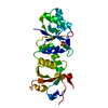
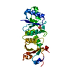
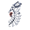
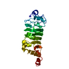
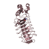
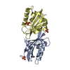
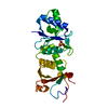
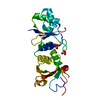
| Title | Lysine-ligated cytochrome c with F82H | ||||||
 Components Components | Cytochrome c iso-1 | ||||||
 Keywords Keywords |  ELECTRON TRANSPORT ELECTRON TRANSPORT | ||||||
| Function / homology |  Function and homology information Function and homology informationRelease of apoptotic factors from the mitochondria / Pyroptosis / Detoxification of Reactive Oxygen Species / Respiratory electron transport / mitochondrial electron transport, cytochrome c to oxygen / mitochondrial electron transport, ubiquinol to cytochrome c / respirasome / mitochondrial intermembrane space / electron transfer activity / heme binding ...Release of apoptotic factors from the mitochondria / Pyroptosis / Detoxification of Reactive Oxygen Species / Respiratory electron transport / mitochondrial electron transport, cytochrome c to oxygen / mitochondrial electron transport, ubiquinol to cytochrome c / respirasome / mitochondrial intermembrane space / electron transfer activity / heme binding / mitochondrion / metal ion bindingSimilarity search - Function | ||||||
| Biological species |  Saccharomyces cerevisiae (brewer's yeast) Saccharomyces cerevisiae (brewer's yeast) | ||||||
| Method | X-RAY DIFFRACTION / SYNCHROTRON / MOLECULAR REPLACEMENT / Resolution: 2.103 Å | ||||||
 Authors Authors | Amacher, J.F. / Zhu, M.Q. / Zhong, F. / Pletneva, E.K. / Madden, D.R. | ||||||
 Citation Citation | Journal: Thesis / Year: 2014 Title: Understanding PDZ Affinity and Selectivity: All Residues Considered Authors: Amacher, J.F. | ||||||
| History |
|
- Structure visualization
| Structure viewer | Molecule: MolmilJmol/JSmol |
|---|
- Downloads & links
-Download
| PDBx/mmCIF format | 4qao.cif.gz | 79.8 KB | Display | PDBx/mmCIF format |
|---|---|---|---|---|
| PDB format | pdb4qao.ent.gz | 60.5 KB | Display | PDB format |
| PDBx/mmJSON format | 4qao.json.gz | Tree view | PDBx/mmJSON format | |
| Others |  Other downloads Other downloads |
-Validation report
| Arichive directory | https://data.pdbj.org/pub/pdb/validation_reports/qa/4qaoftp://data.pdbj.org/pub/pdb/validation_reports/qa/4qao | HTTPS FTP |
|---|
-Related structure data
| Related structure data | 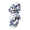 4q5pS S: Starting model for refinement |
|---|---|
| Similar structure data |

-Links
 PDBj
PDBj


- Assembly
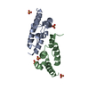
| Deposited unit | 
| ||||||||
|---|---|---|---|---|---|---|---|---|---|
| 1 | 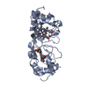
| ||||||||
| 2 | 
| ||||||||
| 3 | 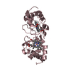
| ||||||||
| Unit cell |
|
-Components
| #1: Protein | Mass: 11690.332 Da / Num. of mol.: 3 / Fragment: UNP residues 8-109 / Mutation: T78C, K79G, F82H Source method: isolated from a genetically manipulated source Source: (gene. exp.) Saccharomyces cerevisiae (brewer's yeast)Gene: CYC1, YJR048W, J1653 / Production host:  Escherichia coli (E. coli) / References: UniProt: P00044 Escherichia coli (E. coli) / References: UniProt: P00044#2: Chemical | Heme B  Mass: 616.487 Da / Num. of mol.: 3 / Source method: obtained synthetically / Formula: C34H32FeN4O4 Mass: 616.487 Da / Num. of mol.: 3 / Source method: obtained synthetically / Formula: C34H32FeN4O4#3: Water | ChemComp-HOH / | Water Mass: 18.015 Da / Num. of mol.: 94 / Source method: isolated from a natural source / Formula: H2O Mass: 18.015 Da / Num. of mol.: 94 / Source method: isolated from a natural source / Formula: H2O |
|---|
-Experimental details
-Experiment
| Experiment | Method: X-RAY DIFFRACTION / Number of used crystals: 1 |
|---|
- Sample preparation
| Crystal | Density Matthews: 3.11 Å3/Da / Density % sol: 60.42 % |
|---|---|
| Crystal grow | Temperature: 291 K / Method: vapor diffusion, hanging drop / pH: 8 Details: 28% w/v PEG, 0.3 M ammonium phosphate, 0.1 M Tris, pH 8.0, VAPOR DIFFUSION, HANGING DROP, temperature 291K |
-Data collection
| Diffraction | Mean temperature: 100 K | ||||||||||||||||||||||||||||||||||||||||||
|---|---|---|---|---|---|---|---|---|---|---|---|---|---|---|---|---|---|---|---|---|---|---|---|---|---|---|---|---|---|---|---|---|---|---|---|---|---|---|---|---|---|---|---|
| Diffraction source | Source: SYNCHROTRON / Site: NSLS  / Beamline: X6A / Wavelength: 1.0781 Å / Beamline: X6A / Wavelength: 1.0781 Å | ||||||||||||||||||||||||||||||||||||||||||
| Detector | Type: ADSC QUANTUM 270 / Detector: CCD / Date: Nov 11, 2013 | ||||||||||||||||||||||||||||||||||||||||||
| Radiation | Monochromator: channel cut Si(111) / Protocol: SINGLE WAVELENGTH / Monochromatic (M) / Laue (L): M / Scattering type: x-ray | ||||||||||||||||||||||||||||||||||||||||||
| Radiation wavelength | Wavelength: 1.0781 Å / Relative weight: 1 | ||||||||||||||||||||||||||||||||||||||||||
| Reflection twin |
| ||||||||||||||||||||||||||||||||||||||||||
| Reflection | Resolution: 2.103→19.52 Å / Num. all: 25279 / Num. obs: 25155 / % possible obs: 99.5 % / Observed criterion σ(F): 1.36 / Observed criterion σ(I): 8.48 / Redundancy: 29.1 % / Rsym value: 0.095 / Net I/σ(I): 39.39 | ||||||||||||||||||||||||||||||||||||||||||
| Reflection shell |
|
- Processing
| Software |
| ||||||||||||||||||||||||||||||||||||||||||||||||||||||||||||||||||||||||||||||||||||||||||||||||||||||||||||||||||||||||||||||||||||||||||||||||||||||||||||||||||||||||||||||||||||||
|---|---|---|---|---|---|---|---|---|---|---|---|---|---|---|---|---|---|---|---|---|---|---|---|---|---|---|---|---|---|---|---|---|---|---|---|---|---|---|---|---|---|---|---|---|---|---|---|---|---|---|---|---|---|---|---|---|---|---|---|---|---|---|---|---|---|---|---|---|---|---|---|---|---|---|---|---|---|---|---|---|---|---|---|---|---|---|---|---|---|---|---|---|---|---|---|---|---|---|---|---|---|---|---|---|---|---|---|---|---|---|---|---|---|---|---|---|---|---|---|---|---|---|---|---|---|---|---|---|---|---|---|---|---|---|---|---|---|---|---|---|---|---|---|---|---|---|---|---|---|---|---|---|---|---|---|---|---|---|---|---|---|---|---|---|---|---|---|---|---|---|---|---|---|---|---|---|---|---|---|---|---|---|---|
| Refinement | Method to determine structure: MOLECULAR REPLACEMENT Starting model: PDB ENTRY 4Q5P Resolution: 2.103→19.52 Å / Cor.coef. Fo:Fc: 0.942 / Cor.coef. Fo:Fc free: 0.915 / SU B: 3.939 / SU ML: 0.113 / Cross valid method: THROUGHOUT / ESU R: 0.037 / ESU R Free: 0.034 / Stereochemistry target values: MAXIMUM LIKELIHOOD / Details: HYDROGENS HAVE BEEN ADDED IN THE RIDING POSITIONS
| ||||||||||||||||||||||||||||||||||||||||||||||||||||||||||||||||||||||||||||||||||||||||||||||||||||||||||||||||||||||||||||||||||||||||||||||||||||||||||||||||||||||||||||||||||||||
| Solvent computation | Ion probe radii: 0.8 Å / Shrinkage radii: 0.8 Å / VDW probe radii: 1.2 Å / Solvent model: MASK | ||||||||||||||||||||||||||||||||||||||||||||||||||||||||||||||||||||||||||||||||||||||||||||||||||||||||||||||||||||||||||||||||||||||||||||||||||||||||||||||||||||||||||||||||||||||
| Displacement parameters | Biso mean: 30.737 Å2
| ||||||||||||||||||||||||||||||||||||||||||||||||||||||||||||||||||||||||||||||||||||||||||||||||||||||||||||||||||||||||||||||||||||||||||||||||||||||||||||||||||||||||||||||||||||||
| Refinement step | Cycle: LAST / Resolution: 2.103→19.52 Å
| ||||||||||||||||||||||||||||||||||||||||||||||||||||||||||||||||||||||||||||||||||||||||||||||||||||||||||||||||||||||||||||||||||||||||||||||||||||||||||||||||||||||||||||||||||||||
| Refine LS restraints |
|