 Movie
Movie Controller

[English] 日本語
 Yorodumi
- PDB-4q94: human RPRD1B CID in complex with a RPB1-CTD derived Ser2 phosphor... -
+ Open data

- Basic information
| Entry | Database: PDB / ID: 4q94 | ||||||
|---|---|---|---|---|---|---|---|
| Title | human RPRD1B CID in complex with a RPB1-CTD derived Ser2 phosphorylated peptide | ||||||
 Components Components |
| ||||||
 Keywords Keywords | PEPTIDE BINDING PROTEIN /  Structural Genomics / Structural Genomics Consortium / SGC Structural Genomics / Structural Genomics Consortium / SGC | ||||||
| Function / homology |  Function and homology information Function and homology informationregulation of cell cycle process / RNA polymerase II promoter clearance / mRNA 3'-end processing / transcription preinitiation complex / RNA polymerase II complex binding / RNA polymerase II C-terminal domain binding / RNA polymerase II transcribes snRNA genes / positive regulation of cell population proliferation / positive regulation of transcription by RNA polymerase II / nucleoplasm ...regulation of cell cycle process / RNA polymerase II promoter clearance / mRNA 3'-end processing / transcription preinitiation complex / RNA polymerase II complex binding / RNA polymerase II C-terminal domain binding / RNA polymerase II transcribes snRNA genes / positive regulation of cell population proliferation / positive regulation of transcription by RNA polymerase II / nucleoplasm / identical protein binding / nucleusSimilarity search - Function | ||||||
| Biological species |  Homo sapiens (human) Homo sapiens (human) | ||||||
| Method | X-RAY DIFFRACTION / SYNCHROTRON / MOLECULAR REPLACEMENT / molecular replacement / Resolution: 1.85 Å | ||||||
 Authors Authors | Ni, Z. / Xu, C. / Tempel, W. / El Bakkouri, M. / Loppnau, P. / Bountra, C. / Arrowsmith, C.H. / Edwards, A.M. / Min, J. / Greenblatt, J.F. / Structural Genomics Consortium (SGC) | ||||||
 Citation Citation | Journal: Nat.Struct.Mol.Biol. / Year: 2014 Title: RPRD1A and RPRD1B are human RNA polymerase II C-terminal domain scaffolds for Ser5 dephosphorylation. Authors: Ni, Z. / Xu, C. / Guo, X. / Hunter, G.O. / Kuznetsova, O.V. / Tempel, W. / Marcon, E. / Zhong, G. / Guo, H. / Kuo, W.H. / Li, J. / Young, P. / Olsen, J.B. / Wan, C. / Loppnau, P. / El ...Authors: Ni, Z. / Xu, C. / Guo, X. / Hunter, G.O. / Kuznetsova, O.V. / Tempel, W. / Marcon, E. / Zhong, G. / Guo, H. / Kuo, W.H. / Li, J. / Young, P. / Olsen, J.B. / Wan, C. / Loppnau, P. / El Bakkouri, M. / Senisterra, G.A. / He, H. / Huang, H. / Sidhu, S.S. / Emili, A. / Murphy, S. / Mosley, A.L. / Arrowsmith, C.H. / Min, J. / Greenblatt, J.F. | ||||||
| History |
|
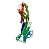
- Structure visualization
| Structure viewer | Molecule: MolmilJmol/JSmol |
|---|
- Downloads & links
-Download
| PDBx/mmCIF format | 4q94.cif.gz | 73.7 KB | Display | PDBx/mmCIF format |
|---|---|---|---|---|
| PDB format | pdb4q94.ent.gz | 53.6 KB | Display | PDB format |
| PDBx/mmJSON format | 4q94.json.gz | Tree view | PDBx/mmJSON format | |
| Others |  Other downloads Other downloads |
-Validation report
| Arichive directory | https://data.pdbj.org/pub/pdb/validation_reports/q9/4q94ftp://data.pdbj.org/pub/pdb/validation_reports/q9/4q94 | HTTPS FTP |
|---|
-Related structure data
| Related structure data | 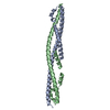 4flaC 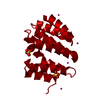 4flbC  4jxtC 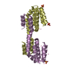 4q96C  4fld C: citing same article ( S: Starting model for refinement |
|---|---|
| Similar structure data |


-Links
 PDBj
PDBj


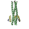
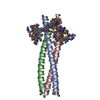
- Assembly
| Deposited unit | 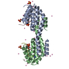
| ||||||||
|---|---|---|---|---|---|---|---|---|---|
| 1 | 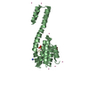
| ||||||||
| 2 | 
| ||||||||
| 3 |
| ||||||||
| Unit cell |
|
-Components
| #1: Protein | Mass: 15560.666 Da / Num. of mol.: 2 Source method: isolated from a genetically manipulated source Source: (gene. exp.) Homo sapiens (human) / Gene: RPRD1B, C20orf77, CREPT / Plasmid: pET15-MHL / Production host:  Escherichia coli (E. coli) / Strain (production host): BL21-V2R-pRARE2 / References: UniProt: Q9NQG5 Escherichia coli (E. coli) / Strain (production host): BL21-V2R-pRARE2 / References: UniProt: Q9NQG5#2: Protein/peptide | Mass: 2363.280 Da / Num. of mol.: 2 / Source method: obtained synthetically / Details: synthetic peptide #3: Chemical | Sulfate  Mass: 96.063 Da / Num. of mol.: 3 / Source method: obtained synthetically / Formula: SO4 Mass: 96.063 Da / Num. of mol.: 3 / Source method: obtained synthetically / Formula: SO4#4: Chemical | ChemComp-UNX /   Num. of mol.: 19 / Source method: obtained synthetically Num. of mol.: 19 / Source method: obtained synthetically#5: Water | ChemComp-HOH / | Water Mass: 18.015 Da / Num. of mol.: 82 / Source method: isolated from a natural source / Formula: H2O Mass: 18.015 Da / Num. of mol.: 82 / Source method: isolated from a natural source / Formula: H2O |
|---|
-Experimental details
-Experiment
| Experiment | Method: X-RAY DIFFRACTION / Number of used crystals: 1 |
|---|
- Sample preparation
| Crystal | Density Matthews: 2.1 Å3/Da / Density % sol: 41.46 % |
|---|---|
| Crystal grow | Temperature: 291 K / Method: vapor diffusion Details: 2.0 M ammonium sulfate, 5% isopropanol, vapor diffusion, temperature 291K |
-Data collection
| Diffraction | Mean temperature: 100 K | |||||||||||||||||||||
|---|---|---|---|---|---|---|---|---|---|---|---|---|---|---|---|---|---|---|---|---|---|---|
| Diffraction source | Source: SYNCHROTRON / Site: APS  / Beamline: 19-ID / Wavelength: 0.97926 Å / Beamline: 19-ID / Wavelength: 0.97926 Å | |||||||||||||||||||||
| Detector | Type: ADSC QUANTUM 315r / Detector: CCD / Date: Jul 18, 2012 | |||||||||||||||||||||
| Radiation | Protocol: SINGLE WAVELENGTH / Monochromatic (M) / Laue (L): M / Scattering type: x-ray | |||||||||||||||||||||
| Radiation wavelength | Wavelength: 0.97926 Å / Relative weight: 1 | |||||||||||||||||||||
| Reflection | Resolution: 1.85→43.11 Å / Num. obs: 26622 / % possible obs: 100 % / Redundancy: 7.1 % / Rmerge(I) obs: 0.057 / Net I/σ(I): 21.7 | |||||||||||||||||||||
| Reflection shell | Diffraction-ID: 1
|
-Phasing
| Phasing | Method: molecular replacement |
|---|
- Processing
| Software |
| ||||||||||||||||||||||||||||||||||||||||||||||||||||||||||||
|---|---|---|---|---|---|---|---|---|---|---|---|---|---|---|---|---|---|---|---|---|---|---|---|---|---|---|---|---|---|---|---|---|---|---|---|---|---|---|---|---|---|---|---|---|---|---|---|---|---|---|---|---|---|---|---|---|---|---|---|---|---|
| Refinement | Method to determine structure: MOLECULAR REPLACEMENT Starting model: pdb entry 4fld 4fld Resolution: 1.85→43.11 Å / Cor.coef. Fo:Fc: 0.963 / Cor.coef. Fo:Fc free: 0.946 / WRfactor Rfree: 0.2161 / WRfactor Rwork: 0.1704 / Occupancy max: 1 / Occupancy min: 0.3 / FOM work R set: 0.8488 / SU B: 2.997 / SU ML: 0.09 / SU R Cruickshank DPI: 0.1294 / SU Rfree: 0.1274 / Cross valid method: THROUGHOUT / σ(F): 0 / ESU R: 0.129 / ESU R Free: 0.127 / Stereochemistry target values: MAXIMUM LIKELIHOOD Details: COOT was used for interactive model building. Model geometry was evaluated with MOLPROBITY.
| ||||||||||||||||||||||||||||||||||||||||||||||||||||||||||||
| Solvent computation | Ion probe radii: 0.8 Å / Shrinkage radii: 0.8 Å / VDW probe radii: 1.2 Å / Solvent model: MASK | ||||||||||||||||||||||||||||||||||||||||||||||||||||||||||||
| Displacement parameters | Biso max: 91.35 Å2 / Biso mean: 31.8293 Å2 / Biso min: 16.02 Å2
| ||||||||||||||||||||||||||||||||||||||||||||||||||||||||||||
| Refinement step | Cycle: LAST / Resolution: 1.85→43.11 Å
| ||||||||||||||||||||||||||||||||||||||||||||||||||||||||||||
| Refine LS restraints |
| ||||||||||||||||||||||||||||||||||||||||||||||||||||||||||||
| LS refinement shell | Resolution: 1.85→1.898 Å / Total num. of bins used: 20
|
